Sporadic bovine lymphoma in calves
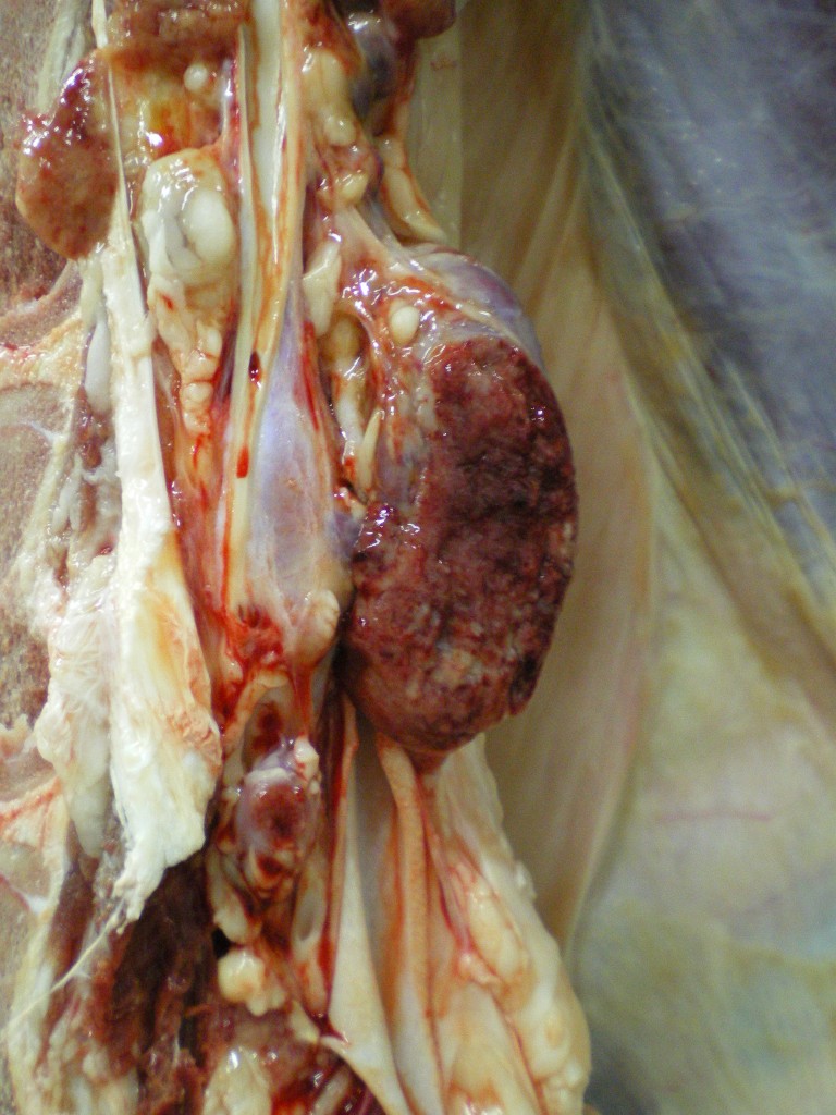
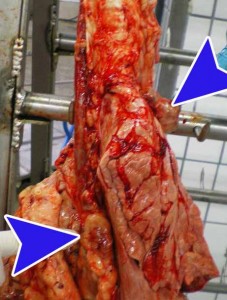
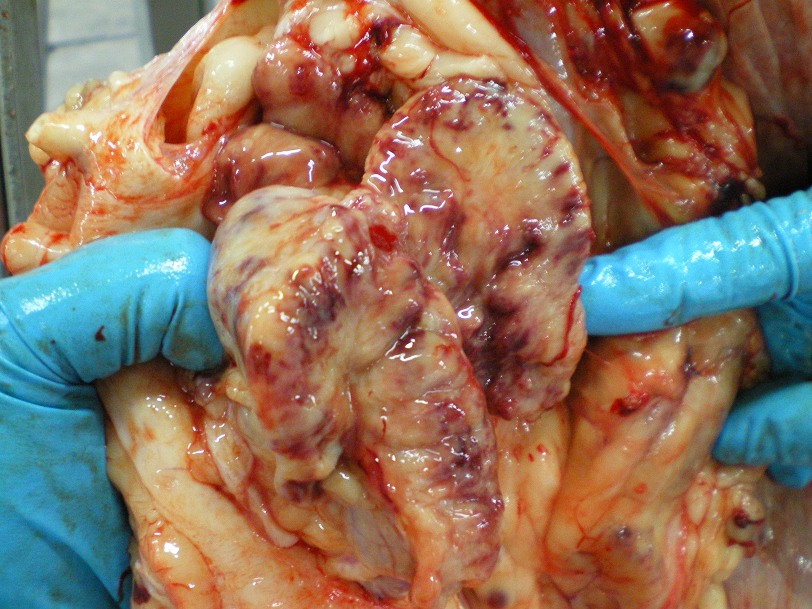
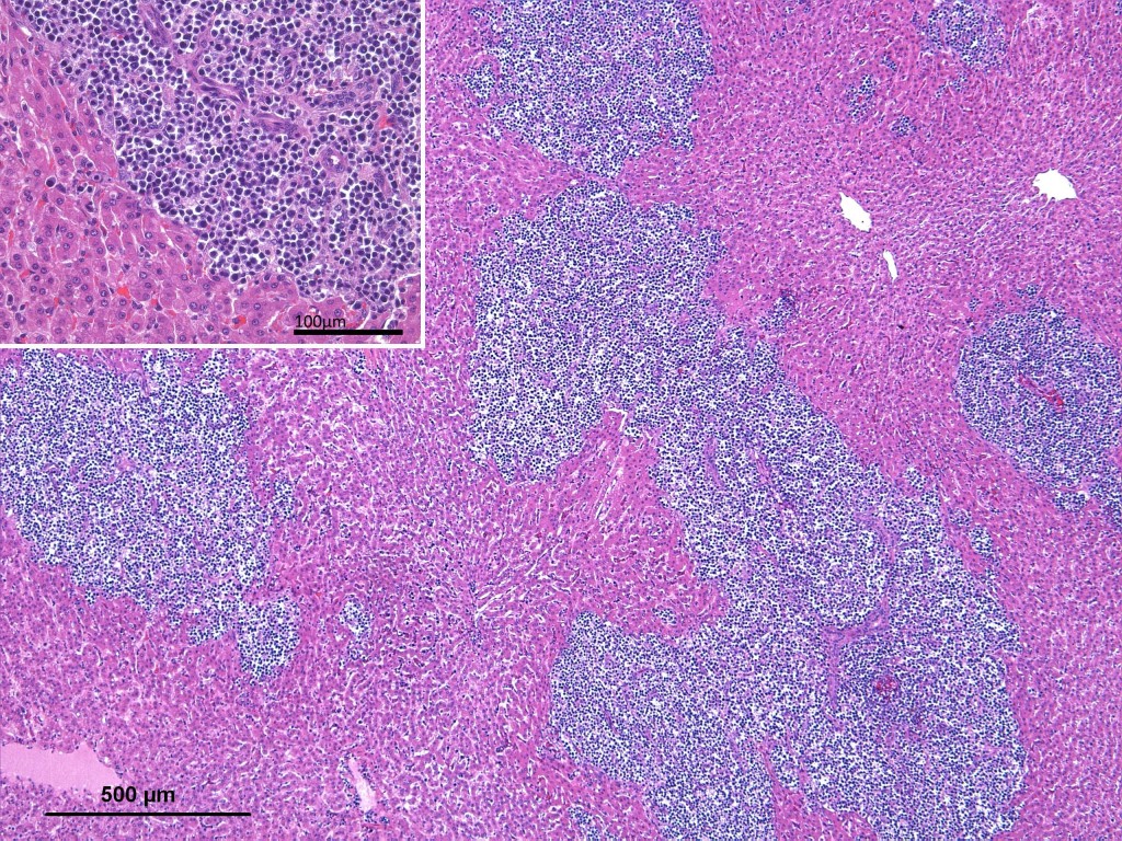
The pathological examination revealed that the affected organs, including the intercostal fat, were infiltrated by a neoplastic lymphoid proliferation. The diagnosis was sporadic bovine lymphoma.
In cattle three types of sporadic lymphoma (of unknown cause) are described:
- The calf sporadic bovine lymphoma: can appear at birth or around 6 months of age. Characteristically it shows symmetrical generalized lymphadenopathy and leukemia. The kidneys are often affected and the liver, spleen and thymus may be involved or not.
- The juvenile form, which predominantly affects the thymus.
- The cutaneous form, more common in animals about two years old.
In adult cattle (mostly around 3 years old) lymphoma may be caused by the enzootic bovine leukosis virus . It is characterized by the appearance of an enlarged lymph node. It usually affects the liver and spleen (hepatomegaly and splenomegaly) and there is also an enteric form described (usually involving the abomasal mucosa). Neoplastic cells can also infiltrate the nerve sheath and enter the epidural fat, compressing the spinal cord.
In the 80s there were a number of cases of leucosis in Catalonia and Spain, due to the importation of infected animals. It is a disease subject to sanitation in Europe, from where it is virtually eradicated Yet, elsewhere, such as in the United States of America it is an endemic disease.


3 comment(s)
If a healthy calf nursed the colistram from a cow with Lymphoma would she be subject to developing the disease.
Of course!!! Colostal transmission: Through the ingestion of milk or colostrum from an infected cow. This can transmit infected cells (B lymphocytes) to the calf.
That would be the case with bovine leucosis, but the sporadic juvenile lymphoma is not transmissible.