Ronyó poliquisitc en un porc
En un porc de raça híbrida de 6 mesos d’edat s’aprecia una alteració bilateral dels ronyons. Aquests presenten una pèrdua de l’estructura normal, amb una evident atròfia de la medul·la i també, en menor mesura, de l’escorça. El parènquima renal presenta un aspecte microquísitc. Els urèters estan molt dilatats i engruixits.
Histològicament s’observa que el parènquima renal esta ocupat en la seva pràctica totalitat per quists recoberts per un epiteli simple, alguns dels qual contenen estructures glomerulars més o menys atrofiades. A l’interstici s’aprecia fibrosi i un component inflamatori mononuclear lleu. De forma aïllada s’observen restes de parènquima renal amb un aspecte aparentment normal (probablement funcionals).
Les lesions observades apunten a una alteració del desenvolupament renal normal ocasionant estructures microquístiques en gairebé la totalitat de l’escorça renal i una pèrdua important de medul•la. Malgrat que no es pot classificar en tipus coneguts de lesió renal, en el diagnòstic diferencial podem incloure:
- Ronyó poliquístic congènit. Mutacions al gen PKD (polycystic kidney disease) poden donar lloc a aquest tipus de lesió que sol ser hereditària.
- Ronyó poliquístic adquirit, potser com a conseqüència d’una inflamació intersticial cronificada o be degut a altres causes que poden desencadenar l’aparició de quists (hipokalemia, alguns medicaments, etc)
- Displàsia renal (defecte en la formació de les nefrones), també congènita. Però la presència focal de nefrones correctament formades fa que aquesta opció sigui menys probable.
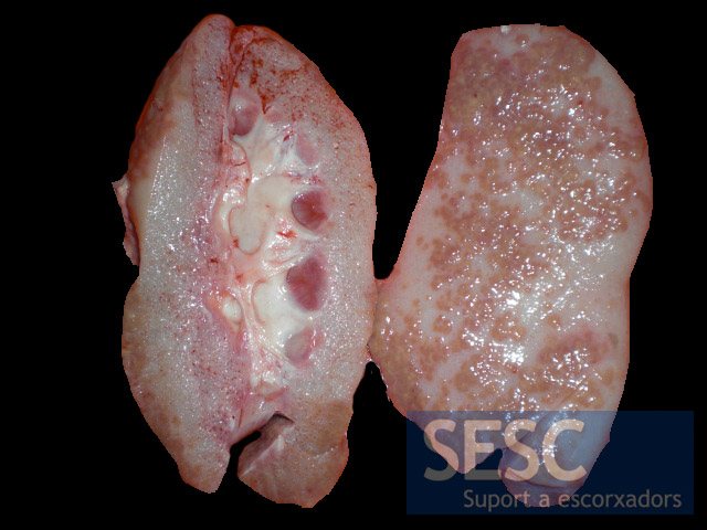
Ronyons pàl•lids, amb irregularitats a la superfície i atròfia de la medul•la i l’escorça. Histològicament, les zones marronoses prominents corresponen a la fracció de parènquima renal “normal” mentre que la resta de parènquima està deprimit i atròfic.

De prop es pot observar una multitud de petits quists que donen un aspecte esponjós al parènquima renal i la manca de medul•la renal.

Microscòpicament, tots els quists estan recobert d’epiteli simple i entre ells s’aprecien restes aïllades de parènquima renal aparentment normal.
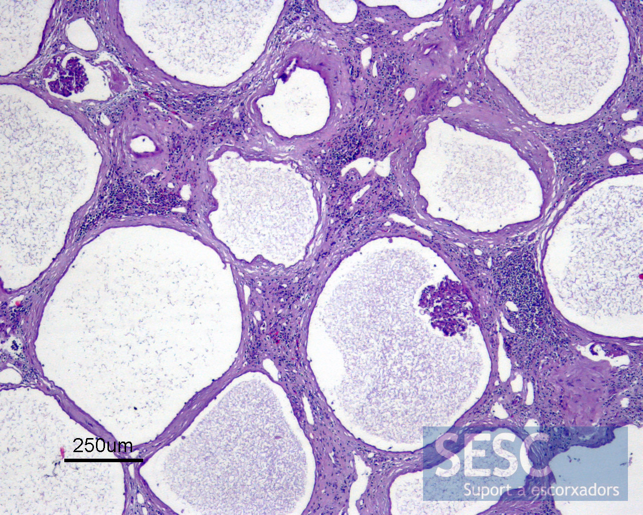
Algunes de les cavitats quístiques contenen un glomèrul renal. L’interstici presenta fibrosi i en algunes zones infiltrat inflamatori mononucelar.

2 comment(s)
Comment from LinkedIn’s Veterinary pathology group from Kendal Harr:
I am posting an article for comparative purposes and because I think it has a good pic of the gross description of poycystic kidney above. Unfortunately, our histo was cut from the article.
Just realized that I cannot attach to this thread
I posted the peer-reviewed pic of polycystic kidneys in color as well as the link to the published article here
https://www.facebook.com/UrikaLLC?ref=hl
Hey, also, please feel free to like the page
Comment from LinkedIn’s Veterinary pathology group from Dr. Lapointe:
Interesting case. Personally I might put renal dysplasia at the top of the differentials list – polycystic kidneys usually show large cysts, sometimes up to 1-2 cm diameter. In this case the cavities are much smaller and appear to mainly be abnormally dilated glomeruli, which could occur from downstream blockage or abnormal development. The presence of normal-looking renal structures does not exclude a diagnosis of renal dysplasia, in which the kidneys are usually a mixture of normal and abnormal nephrons. I would look closely for other histologic features of dysplasia, such as areas of undifferentiated mesenchymal connective tissue, tubules with abnormally hyperplastic lining, glomeruli with fetal appearance, and prominent periglomerular fibrosis.
thanks for sharing the case.