Dermatitis misteriosa
Las lesiones son circulares, de color rojo vivo con coloración marrón en las áreas centrales. Las lesiones son coalescentes y muestran un patrón claramente radial. Desgraciadamente no se pudieron remitir muestras para su análisis laboratorial.
A pesar de la apariencia tan particular de las lesiones no hemos podido determinar cuál es la causa más probable de las mismas. Parece que se trata de una lesión de tipo inflamatorio (dermatitis) pero de etiología desconocida.
Dentro del diagnóstico diferencial se podría incluir:
- Dermatofitosis (tiña): un crecimiento fúngico podría cuadrar con el patrón radial que muestran las lesiones.
- Pitiriasis rosea.
- Mal rojo: a pesar de no observarse la característica morfología romboide de las lesiones.
Cualquiera de estas enfermedades, quizás complicada con infecciones bacterianas secundarias, podrían ser candidatos a tener en cuenta para un posible diagnóstico.
Os invitamos a comentar este caso (al final de la entrada) si habéis visto lesiones similares anteriormente y os animamos a que, si en volveis a verlas, nos hagáis llegar imágenes y muestras para su análisis laboratorial.
Buen verano!
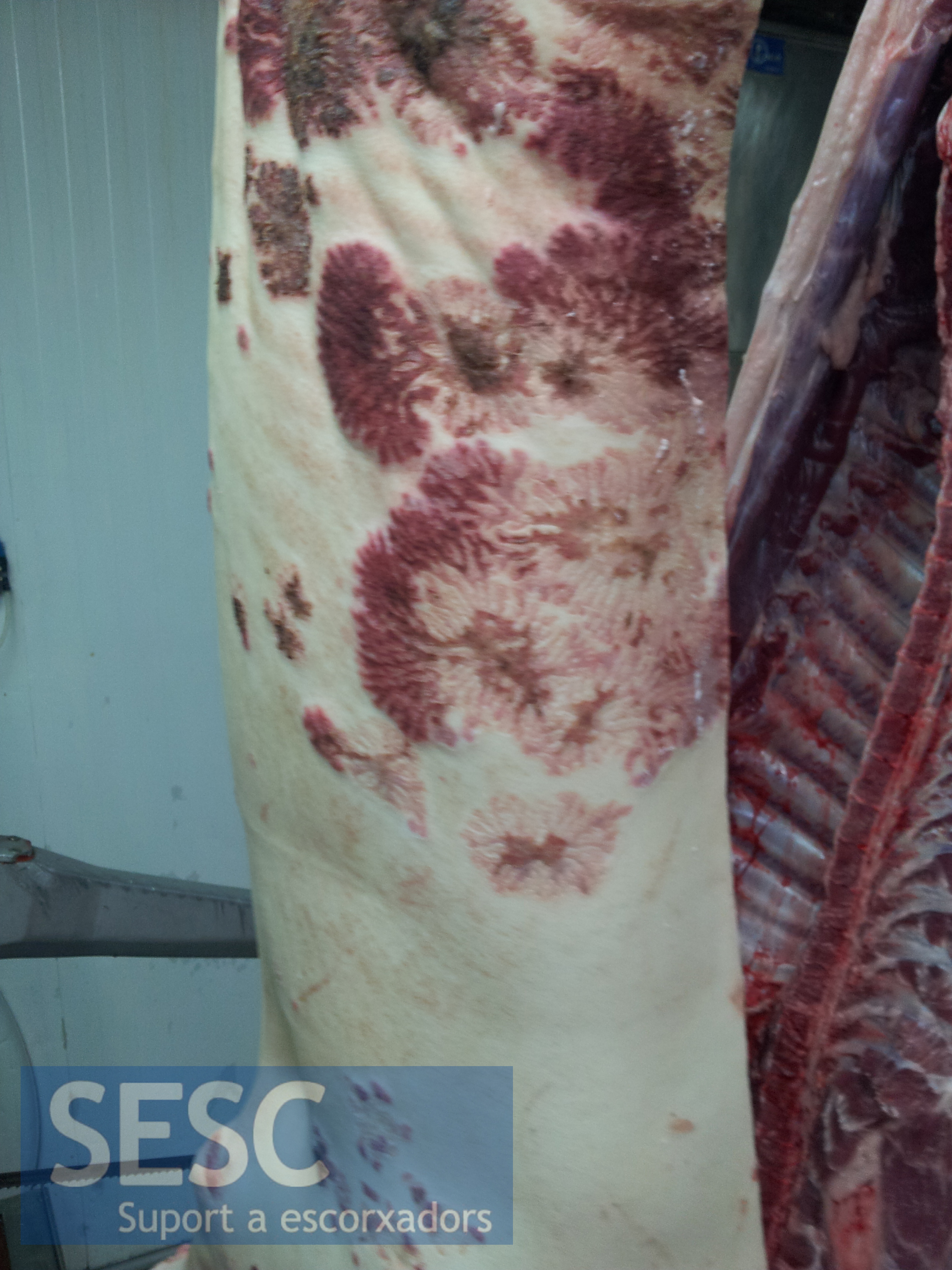
Lesiones multifocales coalescentes conun claro patrón radial.
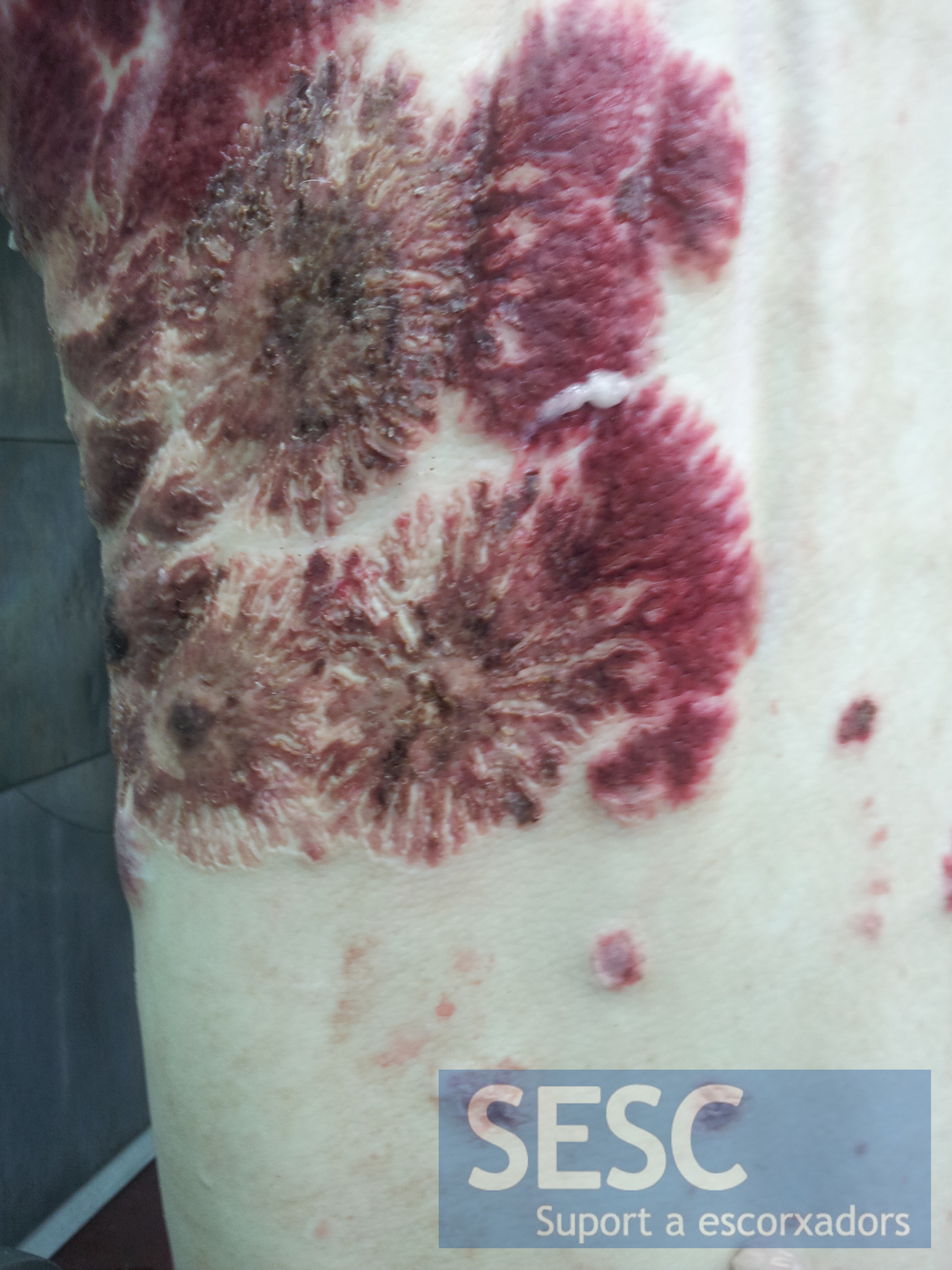
- De más cerca. Lesiones multifocales coalescentes conun claro patrón radial.

4 comment(s)
Here are a few opinions from swine specialist veterinarians in the UK:
1- Think that these lesions are most likely due to Pityriasis Rosea (vegetative dermatitis) also called pustular psoriasiform dermatitis. This is of unknown origin, usually got by Landrace pigs and generally regress spontaneously.
Possible differential diagnosis are Dermatosis vegetans (often associated to pneumonia) and Ringworm but these are much less likely with this presentation.
2- Dramatic lesions. This starburst arrangement I have not seen in pigs before – very strange. I am sure meat inspectors would have mentioned if there was concurrent kidney lesions to support PDNS. However, lots of PDNS cases do not have kidney lesions and also vice versa. Still, I strongly suspect PDNS as being responsible, especially with the central necrosis and surrounding erythema, supporting a vasculitis +/- thrombosis pathogenesis. I definitely have never seen erysipelas with this pattern. Clearly histopath necessary to get a final diagnosis.
3- Another comment was that this could be due to a severe contact dermatitis reaction with a possible vascular component.
4- I suspect the lesions may be the result of a chemical spill with the radiating lesions resulting from spray out from the splashes.
5- Our feeling is that it is probably fungal, given the radiating appearance.
Biopsy with histopath and potentially culture would be the most useful approach. Ideally from a live pig for culture.
6- Thanks for this email – we’ve had a bit of a discussion in the office about these pictures (without knowing any history so we could be completely off the mark!) but it looks vaguely similar to some odd skin lesions we saw with an outbreak of Erysipelas lately. We did wonder with the pictures you’ve sent us whether our lesions would have looked similar given that the pig in the picture has obviously gone through a scald tank/being de-haired at slaughter.
7- Never seen anything quite like this – clearly a slaughter pig so presumably otherwise healthy.
Haemorrhage might suggest possible strange form of PDNS?? Any renal lesions?
8- A strange one! Reminds me of pityriasis rosea perhaps with a secondary fungal infection.
Comment from Swine Health, Nutrition and Production Professionals group in LinkedIn:
I rule out erysipelas and pityriasis rosia. Because these skin diseases have typical lesion which is different from the lesion in your picture. I agree with Dr.Rogerio because the lesion looks like fungal infection. You should do histopathological check and confirm the lesion by fungal laboratory.
It is very interesting lesion. Thank you very much for your sharing.
Comment from Swine Health, Nutrition and Production Professionals group in LinkedIn:
Gross lesions are suggestive of fungal infection or allergic reaction (a component of the diet), followed by secondary infection bactreriana.
For me, Erysipelas is ruled out, for even in an atypical form, would have to be epidemic and not an isolated case, and most other symptoms (hyperthermia, mortality, unvaccinated flock, etc …).
My recommendation is to follow the property, to see if there outtros cases, and in this case, perform the collection of material for a specific diagnosis.
Comment from Veterinary Pathology group in LinkedIn:
This is an interesting case. Sad to know that there’s no sample collected for laboratory workups.