Chronic myocardial infarction in a calf
We receive an inquiry regarding a 10-month-old bovine carcass (Frisian breed), that presented a nodular lesion in the myocardium. Since the presence of muscular nodular lesions in bovine rises the suspicion of cysticercosis, the whole heart was submitted to SESC to rule out such possibility and to reach a diagnosis. Moreover, the carcass was condemned due to having septic arthritis, although this lesion was not studied through SESC.
The myocardial lesion had rounded scction of approximately 1cm in diameter and 3cm in length and was localized in the subepicardial tissue. The lesion was firm and presented a white homogeneous coloration.
At histopathological study, the lesion consisted of a focus of cardiomyocyte necrosis, surrounded by abundant granulation tissue and fibrosis. The cardiomyocytes in the necrotic focus presented karyolisis and karyorrhexis (fragmentation and loss of nuclei), but the cellular morphology was conserved. This change is characteristic of coagulative necrosis, which indicates ischemic damage (focal infarction). Surrounding the necrotic tissue, little amounts of neutrophils and abundant macrophages were seen. These inflammatory cells are drawn the necrotic tissue to phagocytize the necrotic debris in a process called necrotaxis. Finally, the lesion was circumscribed by a wide capsule composed of granulation tissue and mature fibrous tissue, being this a sign of the chronicity of the lesion.
The presence of necrotic tissue under the process of encapsulation is referred to as sequestrum, and it’s the effort of the organism to delimitate and repair the dead tissue. In this case, it is a sign that a focal vascular damage produced the ischemic necrosis of the myocardium (infarction) and that the reparation process had begun. Although many insults can affect the myocardium and, specifically, produce infarction through vascular damage, the presence of a unique focal infarct is not consistent with agents producing primary cardiomyocyte damage. The most probable event is an arterial occlusive process. Although the vessels adjacent to the lesion were examined, no vascular histologic anomaly could be identified. It is possible that the vascular lesion had been resolved by the time of slaughter and that only the infarcted area remained as its consequence. (AC)
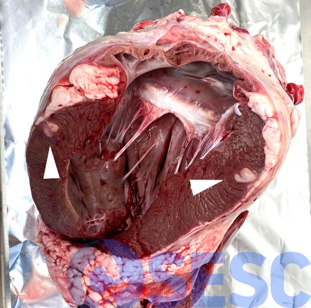
Bovine heart. Once the left ventricle was sectioned, a focal nodulation can be seen in subepicardial localization (arrows).
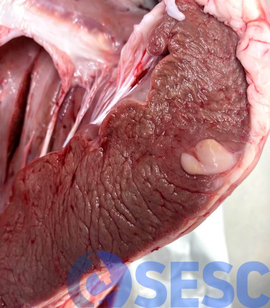
Bovine heart. Detail of the lesion, which shows a homogeneous white coloration.
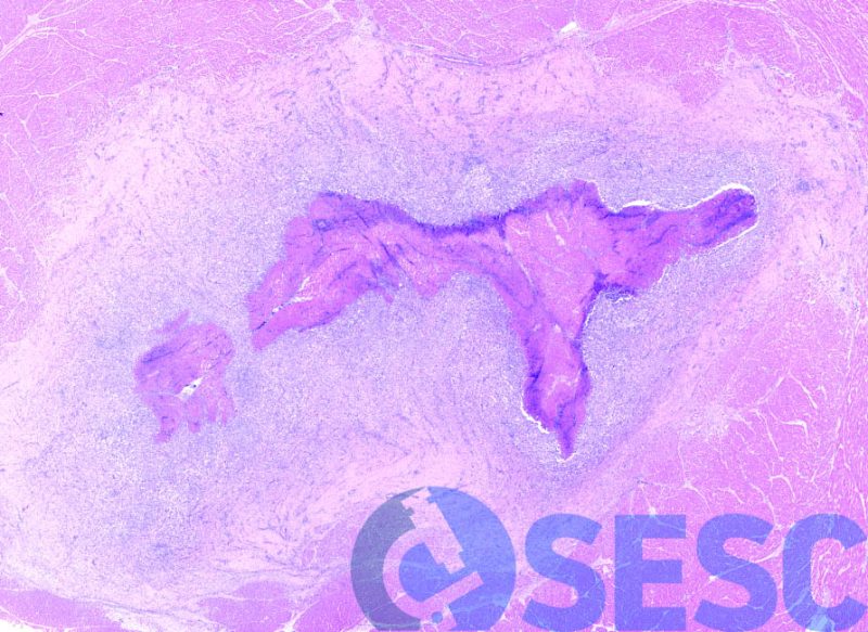
Histologic image at low magnification, with necrotic cardiac tissue in the centre of the lesion surrounded by a wide capsule.
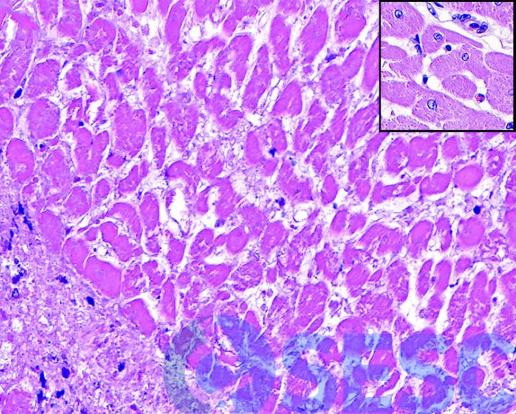
Detail of the necrotic tissue, where the loss of the nuclei of the cardiomyocytes can be observed, especially evident when compared with the adjacent viable tissue (insert).
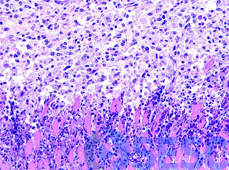
Margin of the necrotic tissue, where neutrophils (bottom half) and macrophages (top) can be seen phagocytizing necrotic debris.
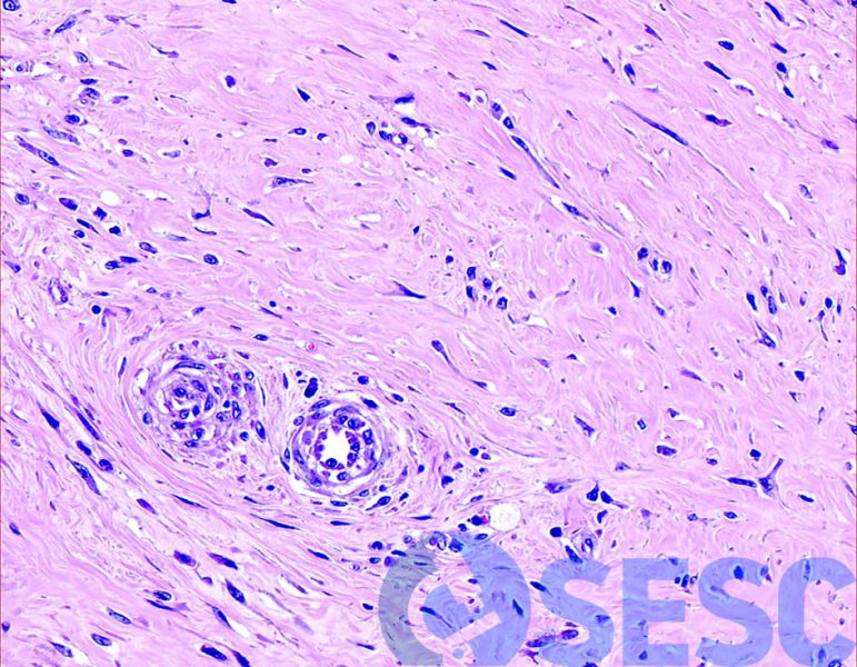
Detail of the capsule in the outermost region, where abundant mature fibrous tissue can be seen with large quantities of collagen, product of its chronicity.
