Hepatic necrobacillosis in a bovine carcass
In a 12-month-old male Friesian calf carcass, a generalized yellowish color change and the presence of multiple nodular lesions in the liver were observed. These were random multifocal lesions between 1-2 cm in diameter scattered throughout the organ, whitish in color and with a peripheral reddish halo, which were also found deep into the parenchyma and had a necrotic (non-exudative) appearance.
Histologically, the liver lesions corresponded to areas of coagulative necrosis surrounded by a halo of a mixed inflammatory reaction, this led to the classification of the lesion as generalized, subacute, multifocal necrotizing hepatitis of probable bacterial origin.
Microbiological culture of a sample of the liver was done to detect anaerobic bacteria and growth of Fusobacterium necrophorum was observed.
It is therefore a case of hepatic necrobacillosis, this lesion is described in calves and lambs as a complication of omphalophlebitis (inflammation of the umbilical cord) or as a complication of ruminitis (inflammation of the rumen due to ruminal acidosis) in cattle. In the latter case, the embolization of the bacteria from the ruminal mucosa, through the portal circulation, accesses the liver, where it spreads and generates the characteristic lesions.
It is not clear that this liver lesion can explain the change in color of the canal due to hepatic jaundice. But Fusobacterium can eliminate toxins with a hemolytic effect and it is highly likely that the animal was septicemic, which could cause pre-hepatic jaundice. (EV)

Calf liver with generalized multifocal, nodular lesions.
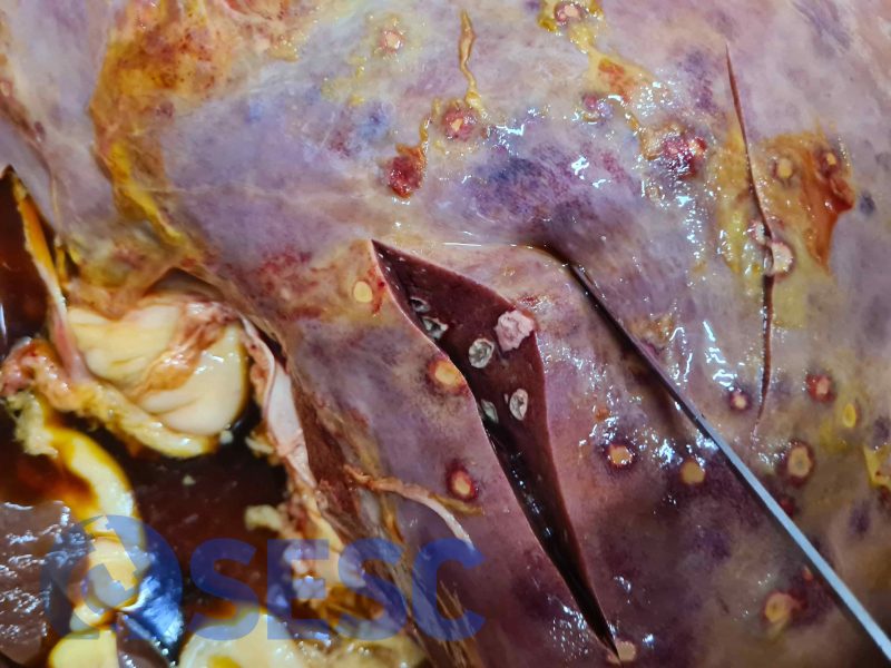
Upon section it can be seen that there are also lesions inside the parenchyma.
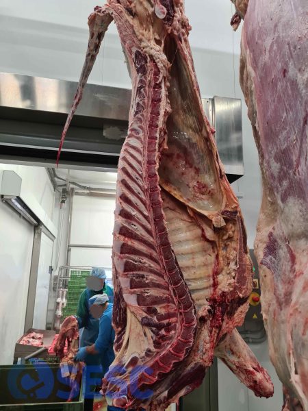
Bovine carcass with slight-moderate yellowish discoloration.
