What is your diagnosis? (141)
At the end of the post you can vote for the diagnosis you think is most correct!
Click on the option that you think is correct and press the "Submit" button to find out if you got it right.
We have received images and samples from a 12-month-old female cross-breed bovine in which firm, nodular lesions 1 cm in diameter are observed throughout the body, but are more frequent on the skin of the neck and upper parts of the forelimbs. (EV)
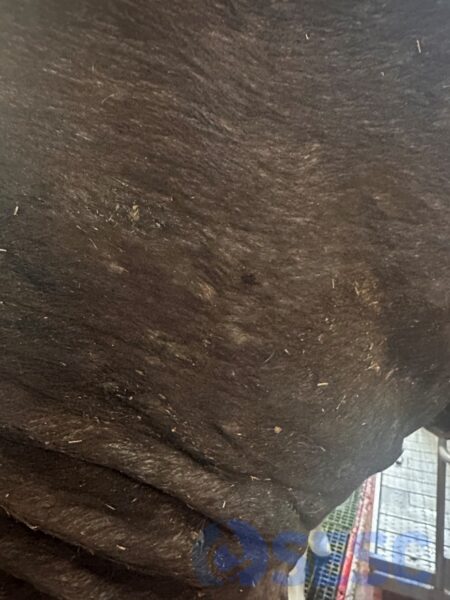
Bovine hide with multiple nodular lesions 1 cm in diameter in the neck region.
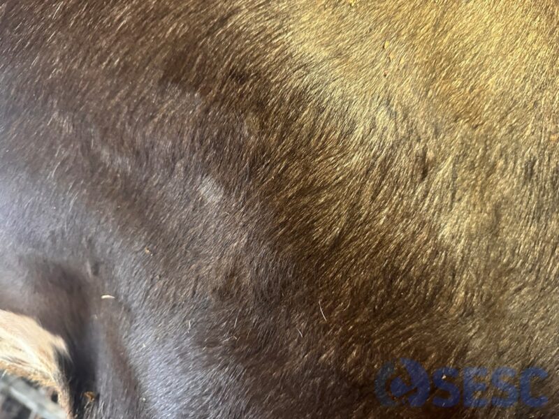
Another image of bovine skin showing multiple nodular lesions 1 cm in diameter. On inspection of the skin fragment sent to us, the nodules were not easily visible; one had to palpate the skin to find them.
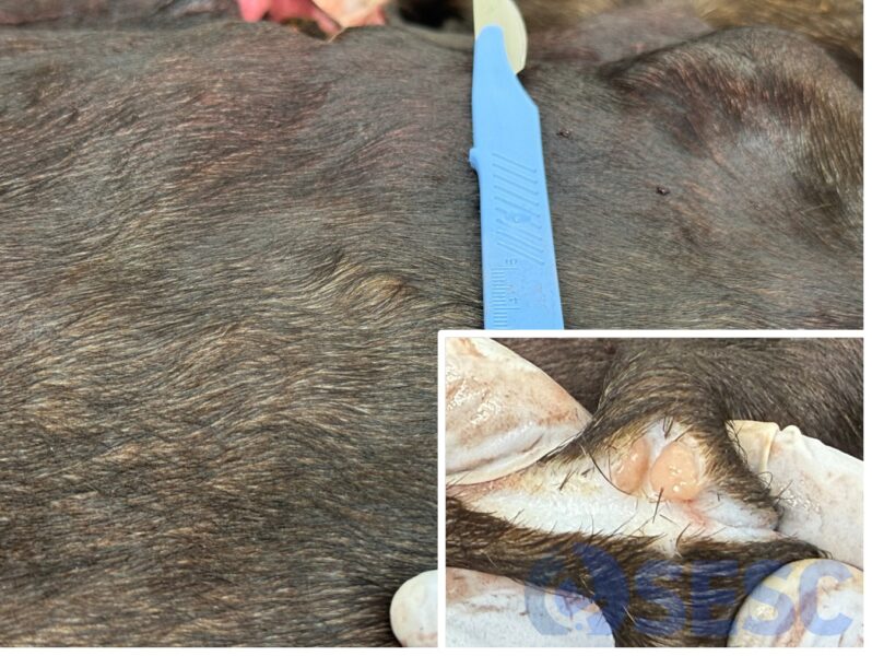
Image of one of the lesions. In the white box, the cream-coloured surface of the lesion in the deep dermis can be seen after being cut open.
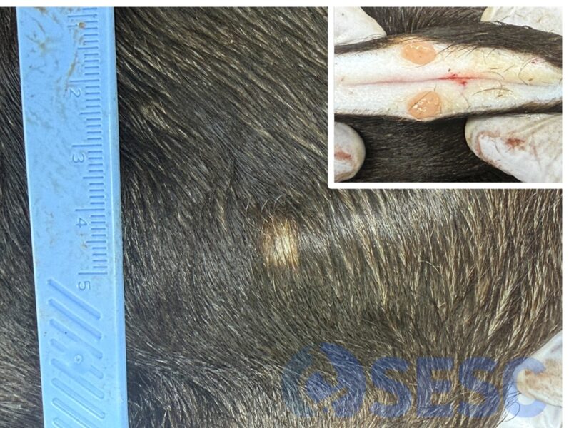
Image of another of the lesions, this one was slightly alopecic. In the white box, the surface of the lesion is visible once cut, looking very similar to the previous one, also located in the deep dermis.
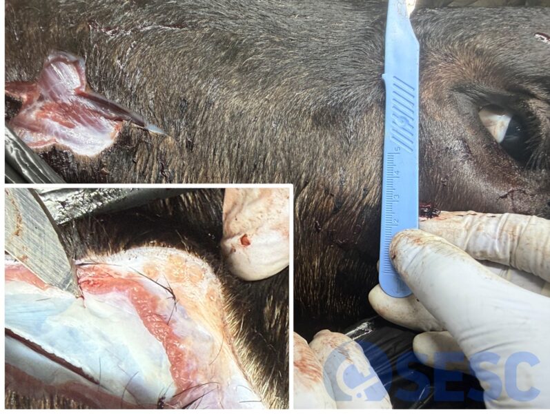
Example of another lesion, located on the head. On section it is observed that it is a multinodular lesion, also confined to the dermis.