Crateriform lesions in the heart of a calf
An inquiry was submitted regarding the heart of a male, Frisian breed, 12 months old calf, presenting hypertrophy of the cardiac atrium with the presence of rounded variable in size and with a crater-like appearance. Additionally, hypertrophy of the wall of the brachiocephalic trunk vessels was observed.
Upon receiving the sample, lesions ranging from 0.3-1.5 cm in diameter were observed, rounded, whitish in color, with a depressed center. Upon section, it was confirmed that these lesions consisted of cystic cavities with a small amount of transparent fluid inside and a whitish wall, 1 mm thick. Additionally, it presented multifocal to coalescent areas, whitish in color, moderately penetrating into the muscle tissue.
A histopathological study was conducted, and it was observed that the lesions corresponded to a proliferation of large caliber blood vessels, located in the epicardium, forming irregular lumens without blood in them. The vessel walls consisted of an endothelium of normal appearance, without mitosis or cellular atypia, a moderately disorganized tunica media with deposits of myxoid basophilic material, elastin fibers in a segmental pattern, and the presence of numerous blood capillaries; and a tunica adventitia, also disorganized, that could occasionally fuse with adjacent vessels. Among these vascular structures, normal and hypertrophied myocytes intermingled with fibrous tissue and adipocytes were frequently seen. In this way, these lesions were classified as vascular hamartomas located in the atrium of the heart.
Cardiac vascular hamartoma consists of an excessive and disorganized proliferation of vascular tissue, and due to its limited growth, it is considered a malformation and not a neoplasm. In veterinary medicine, they have been described in cattle, and the lesions were incidental findings at the slaughterhouse. They differ from a hemangioma since in a hamartoma, all the layers constituting a blood vessel can be found proliferating, whereas hemangiomas are proliferations of only endothelial cells. (MC)
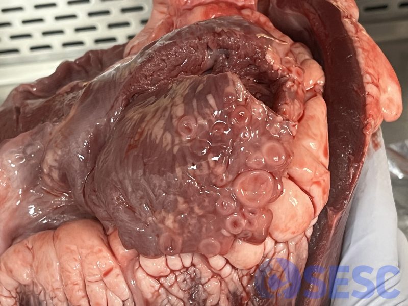
Bovine heart. Rounded, whitish lesions with a depressed center, measuring between 0.3-1.5 cm in diameter, are observed in the atrium of the heart.
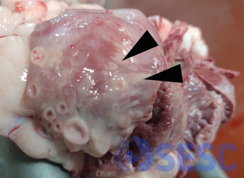
Bovine heart. Whitish, multifocal-coalescent lesions which penetrate the cardiac muscle (black arrows).
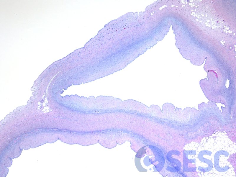
Histological image at low magnification, showing disorganized and anastomosed vascular proliferations, with irregular lumens without the presence of blood, separated by connective tissue and myocardium. Hematoxylin-eosin (HE)
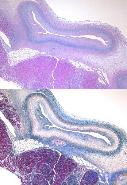
Top image: Hematoxylin-eosin (HE). Bottom image: Masson's trichrome: The proliferating vessels present an inner layer of loose collagen (blue stain), disorganized and intermixed with elastin fibers, and an outer layer of denser collagen. Muscle fibers show an intense pink coloration.
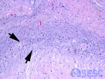
At higher magnifications, in the wall of these vascular structures, cardiomyocytes (black arrows) intermixed with fibrous tissue and adipocytes can be observed.
