19/07/2012
|
Bovine
2
What is your diagnosis? (2)
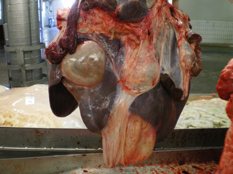
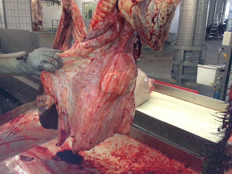
The lesions shown in the images are from bufalos of the same herd.
Microscopically, in both cases, cystic lesions were observed associated to a granulomatous immfalamtory infiltrate, with abundant eosinophils.
ANSWER:
An 80% of the answers were right to establish a diagnosis of a parasitic disease. The intraparenquimatous localization of the cysts pointed to a diagnois of an Hydatid cyst (both E.granulosus and E. multilocularis can produce such lesions).
From a total of 26 answers: 50% E. granulosus, 31% E. multilocularis, 12% Congenital epithelial cysts and 8% C. tenuicuollis.


2 comment(s)
Buenos dias!
Un 81% de las respuestas han acertado que se trata de quistes de orígen parasitario. Su localización intraparenquimatosa és característica de una Hidatidosis (E.granulosus ó E. multiloculris).
De un total de 26 respuestas: 50% E. granulosus, 31% E. multilocularis, 12% Quistes epiteliales congénitos y 8% C. tenuicuollis.
Bon dia!
Com be ha encertat un 81% de les respostes es tracta de quists d’origen parasitari. La seva localització intraparenquimatosa és característica d’una Hidatidosi (E.granulosus o E. multiloculris).
D’un total de 26 respostes: 50% E. granulosus, 31% E. multilocularis, 12% Quists epitelials congènits i 8% C. tenuicuollis.