Neumonía granulomatosa (de causa desconocida) en un cerdo
Microscópicamente estas masas corresponden a un intenso infiltrado inflamatorio formado mayoritariamente por macrófagos, escasos eosinófilos y con presencia de células gigantes multinucleadas. No se aprecia necrosis. Se examinan secciones de linfonodo mediastínico que presentan también inflamación del mismo tipo. Se trata, por tanto, de una neumonía y linfadenitis granulomatosas proliferativas.
Desgraciadamente no se llega a determinar la causa de esta reacción inflamatoria, las pruebas que se han hecho incluyen:
- Se descarta que se trate de una micobacteriosis mediante una tinción de Ziehl-Neelsen (Negativo), una PCR para el complejo M.tuberculosis (Negativo) y un cultivo de micobacterias (Negativo).
- Se descarta la presencia de estructuras fúngicas mediante una tinción de Grocott (Negativo).
- Se descarta la presencia de bacterias gram positivas con una tinción de gram (Negativo) y un cultivo microbiológico estándar (Negativo).
- Morfológicamente, se descarta también que se trate de un proceso asociado a parásitos o una neoplasia.
Si alguien se ha encontrado con casos similares o tiene una idea de qué puede causar este tipo de lesión le/la invitamos a compartirlo en la sección de comentarios que hay al final de esta entrada.
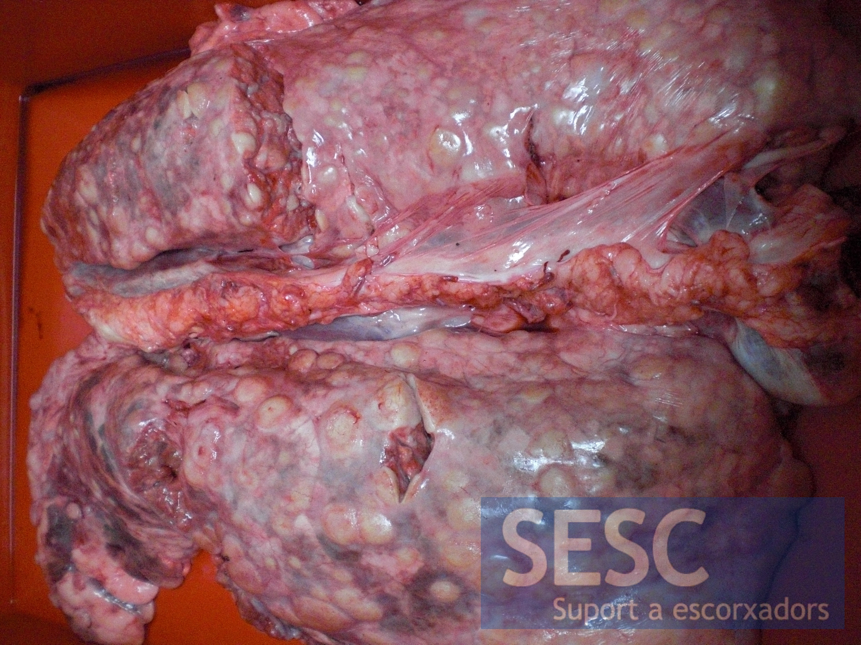
La lesión afecta a todos los lóbulos pulmonares y los linfonodos mediastínicos.
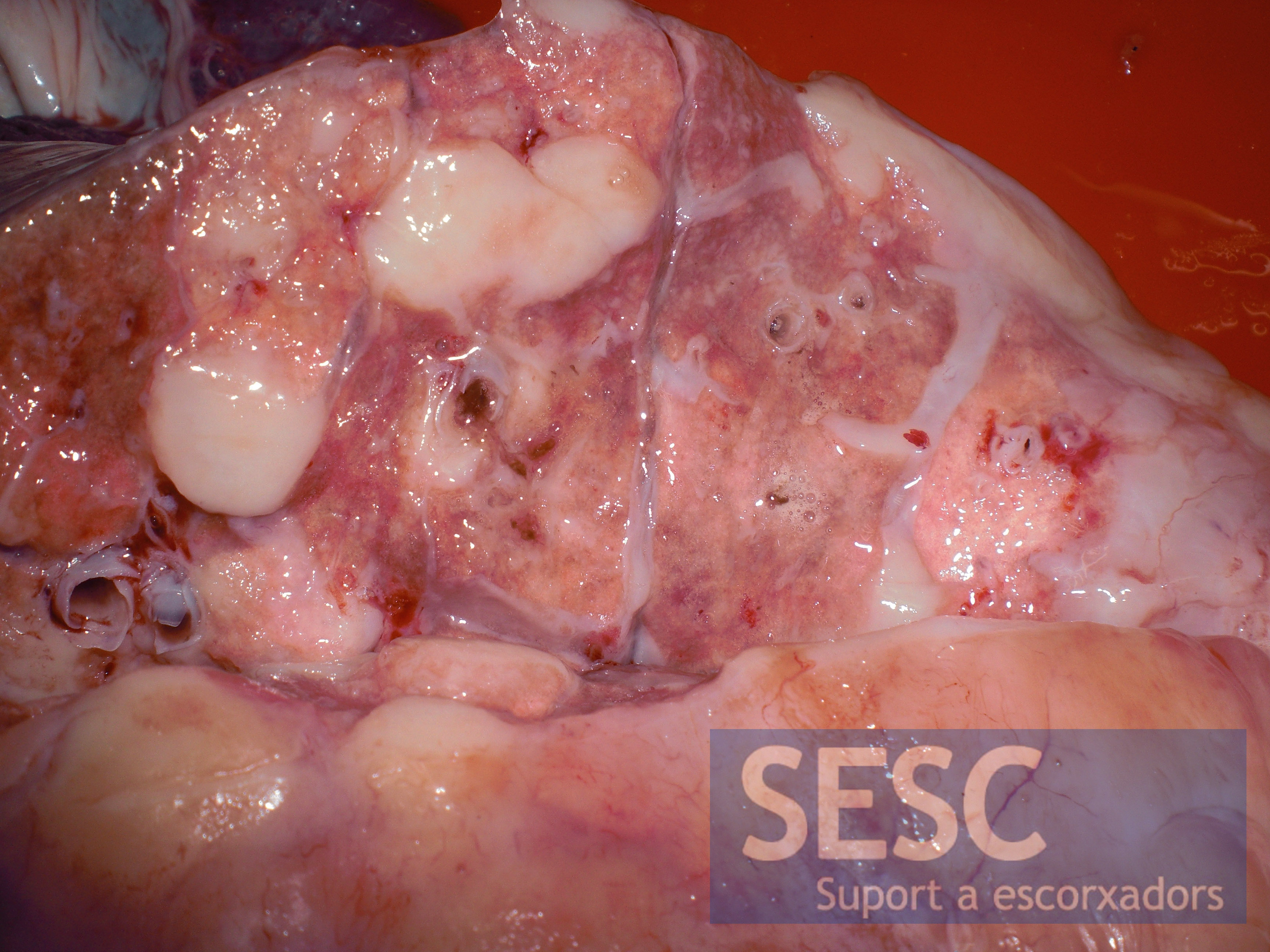
En la sección las masas presentan una coloración blanquecina homogénea.
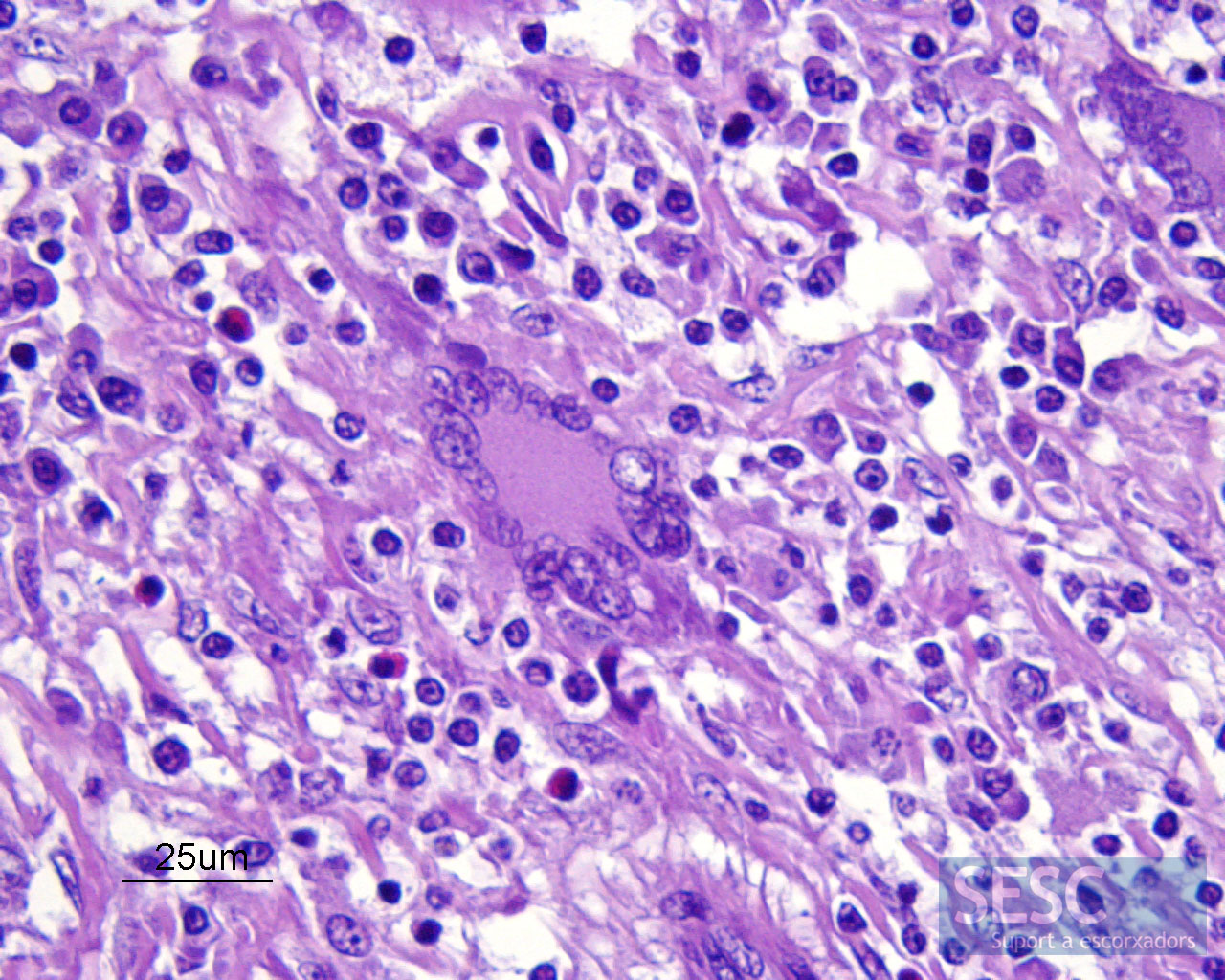
Al microscopio se puede observar una reacción inflamatoria granulomatosa con abundantes células gigantes multinucleadas como las de la imagen.

7 comment(s)
De : aavld-request@ucdavis.edu [mailto:aavld-request@ucdavis.edu] De la part de Glenn Songer
Envoyé : 24 mars 2014 17:17
À : aavld@ucdavis.edu
Objet : Re: Granulomatous pneumonia (of unknown cause) in a pig
This picture from Ted has gross lesions very much like what I saw in M. kansasii all those years ago.
Glenn
De : aavld-request@ucdavis.edu [mailto:aavld-request@ucdavis.edu] De la part de Ted Clark
Envoyé : 24 mars 2014 13:53
À : aavld@ucdavis.edu
Objet : Re: Granulomatous pneumonia (of unknown cause) in a pig
I agree with Glenn and Paco this still could be Mycobacteriosis. Here is a pic of a porcine lung that I received from a slaughterhouse many years ago. The lesions were similar to the case being discussed and after much searching on AF stains (probably both ZN and Fites) I did find some AF bacteria. No cultures done. If this pic does not display properly, someone tell me.
Sorry I don’t have access to the block or slide to take a histo picture for you.
Ted Clark
Calgary
De : aavld-request@ucdavis.edu [mailto:aavld-request@ucdavis.edu] De la part de Glenn Songer
Envoyé : 23 mars 2014 20:24
À : aavld@ucdavis.edu
Objet : Re: Granulomatous pneumonia (of unknown cause) in a pig
It doesn’t seem like a great possibility, but Paco might be right. I’ve seen lesions like you describe in both field cases (which are mainly infections of cervical and mesenteric LN) and in experimental cases (when the person doing the inoculation was a little careless — that was me, by the way) and inoculum was partially swallowed to try to infect LN) and partially inhaled (producing the subject lesions in lungs. The experimental cases occurred with both M. avium ss avium (just M. avium in those days) and M. kansasii. It might be worth digging up the methods for mycobacterial culture (Chuck Thoen at ISU should be a good source).
Glenn
De : aavld-request@ucdavis.edu [mailto:aavld-request@ucdavis.edu] De la part de Ramos-Vara, Jose A
Envoyé : 22 mars 2014 16:38
À : aavld@ucdavis.edu
Objet : RE: Granulomatous pneumonia (of unknown cause) in a pig
Despite not being typical of PCV2 infection, I assume that this animal was tested for with negative results. It resembles hairy vetch eosinohilic granulomatous nephritis in cattle. Only one animal affected in the herd? Apparently Actinobacillus porcitonsillarum can produce similar inflammation but I am not sure if it includes eosinophils. Pepe Ramos
De : aavld-request@ucdavis.edu [mailto:aavld-request@ucdavis.edu] De la part de Fabio Del Piero
Envoyé : 22 mars 2014 14:22
À : aavld@ucdavis.edu
Objet : RE: Granulomatous pneumonia (of unknown cause) in a pig
If it would be my case I would contact Roger Maes at MSU for a generic herpesvirus PCR from the paraffinized lung tissues. In case of positive result I would narrow the search toward gammaherpesviruses.
It is vaguely reminiscent of some gammaherpesvirus associated lesions in horses.
Fabio Del Piero
Professor of Pathology
Louisiana State University
==
Comment from Swine Health, Nutrition and Production Professionals group in LinkedIn:
Rogerio Paulo Tovo
Swanager
Dear SESC went their differential diagnosis , and quick reference to the book Diseases of Swine of the Dr. Leman et al. , recommend researching Yersinia pseudotuberculosis , and very many smaller chance of occurrence for Brucella suis . Surely the case is atypical, disease macroscopia is suggestive of tuberculosis , and to a lesser extent atypical case of Staphylococcus aureus , cases that have been discarded by laboratory tests performed .
The symptoms , if your lordship has access , it is important to suggest some way in these cases .
Good luck in continuing the search , and when you find something , let us know . Hugs , Rogério Tovo – Brazil .
I have not seen porcine lung lesions just like this, but given the negative test results so far and the giant cells present, I would next run IHC for PCV2. You could do PCR for PCV2 as well, but the virus is so ubiquitous a positive result would not confirm it caused these lesions.
I assume you used a polarizer on the lesions looking for aspirated foreign material and crystals? A PAS stain might be worth doing as well.