09/02/2015
|
Oví
2
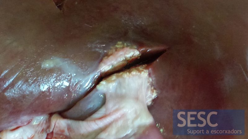
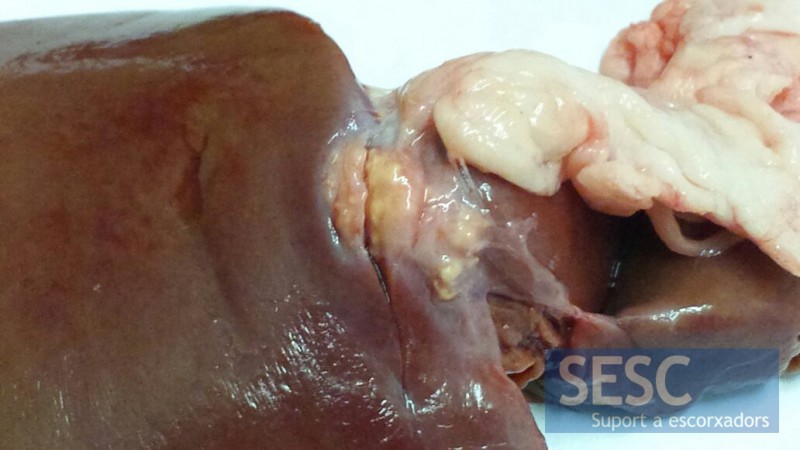
Granulomes hepàtics en fetges de xai
En ambdós casos les lesions eren superficials i presentaven adherències a altres seroses. Histològicament s’apreciaven canvis inflamatoris granulomatosos: extensa necrosi central rodejada d’abundants macròfags epitelioides, alguna cèl·lula gegant multinucleada i presència de leucòcits polimorfonuclears eosinòfils.
Les lesions son suggestives d’una etiologia parasitària i per localització podrien ser compatibles amb un quist de Cysticercus tenuicollis degenerat. En el cas dos es va descartar la presència de micobacteris mitjançant tinció de Ziehl Neelsen, PCR i cultiu de micobacteris ja que la lesió va arribar com una sospita de tuberculosi, malaltia a la que les ovelles també són susceptibles.


2 comment(s)
Comment from Veterinary Pathology group in Linked IN:
By Hala el Miniawy
Vice dean at faculty of veterinary medicine
I think its a pseudo-tuberculosis ovis
Comment from Veterinary Pathology group in Linked IN:
Dave Getzy
Pathologist at IDEXX Laboratories
Hi,
I really appreciate you putting these great cases out for viewing. When I was at the Colorado State University Diagnostic Lab, we used to see these lesions sporadically in lambs, with the same histopathology as you describe. Tuberculosis is always a differential, the eos are usually a good clue, but always a good idea to at least do an acid fast stain to be sure. As much as we like to think one animal- one disease, occasionally, especially in some of the more poorer feedlots, more than one disease may be present at the same time.
Best regards,
Dr. Dave G.
Fort Collins, CO