Follicular chronic lymphoplasmacytic gastritis in a pig
An inspector has submitted a sample of a pig's stomach (6 months old, crossbreed). During the inspection of the viscera at the abattoir, once the stomachs had been cleaned, on exiting the centrifuge, the presence of whitish miliary lesions was observed on the gastric mucosa of one of the stomachs, in the lesser curvature area.
Despite the artefacts caused to the mucosa by the cleaning process, the histopathological examination confirmed that this macroscopic image corresponds to the presence of clusters of lymphocytes and plasma cells forming small follicles in the gastric mucosa. A diagnosis of chronic follicular lymphoplasmacytic gastritis was therefore established.
This finding is non-specific and possibly multifactorial, it could be associated with chronic irritation, dietary factors, old, already healed erosions of the mucosa, an immune-mediated response of unknown aetiology, or the presence of Helicobacter-type bacteria that are very frequently found in the glandular region of pigs' stomachs. However, the relationship between the presence of Helicobacter and the presence of gastric lesions is uncertain, as in the majority of pigs it does not cause lesions. (EV)

Pig's stomach. Milky white miliary lesions on the gastric mucosa.
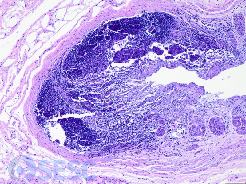
Gastric mucosa with lymphoid cell clusters.