Granulomatous-suppurative pneumonia due to Nocardia in a calf
Histologically an encapsulated lesion was observed with coalescent areas of suppurative necrosis and abundant macrophages. Gram staining allowed to observe the presence of abundant filamentous Gram positive bacteria.
The microbiological culture identified abundant growth of bacteria of the genus Nocardia.
Being an lesion of caseous aspect it was necessary to rule out that it was a case of bovine tuberculosis.

Granulomatous lesion in the lung parenchima.
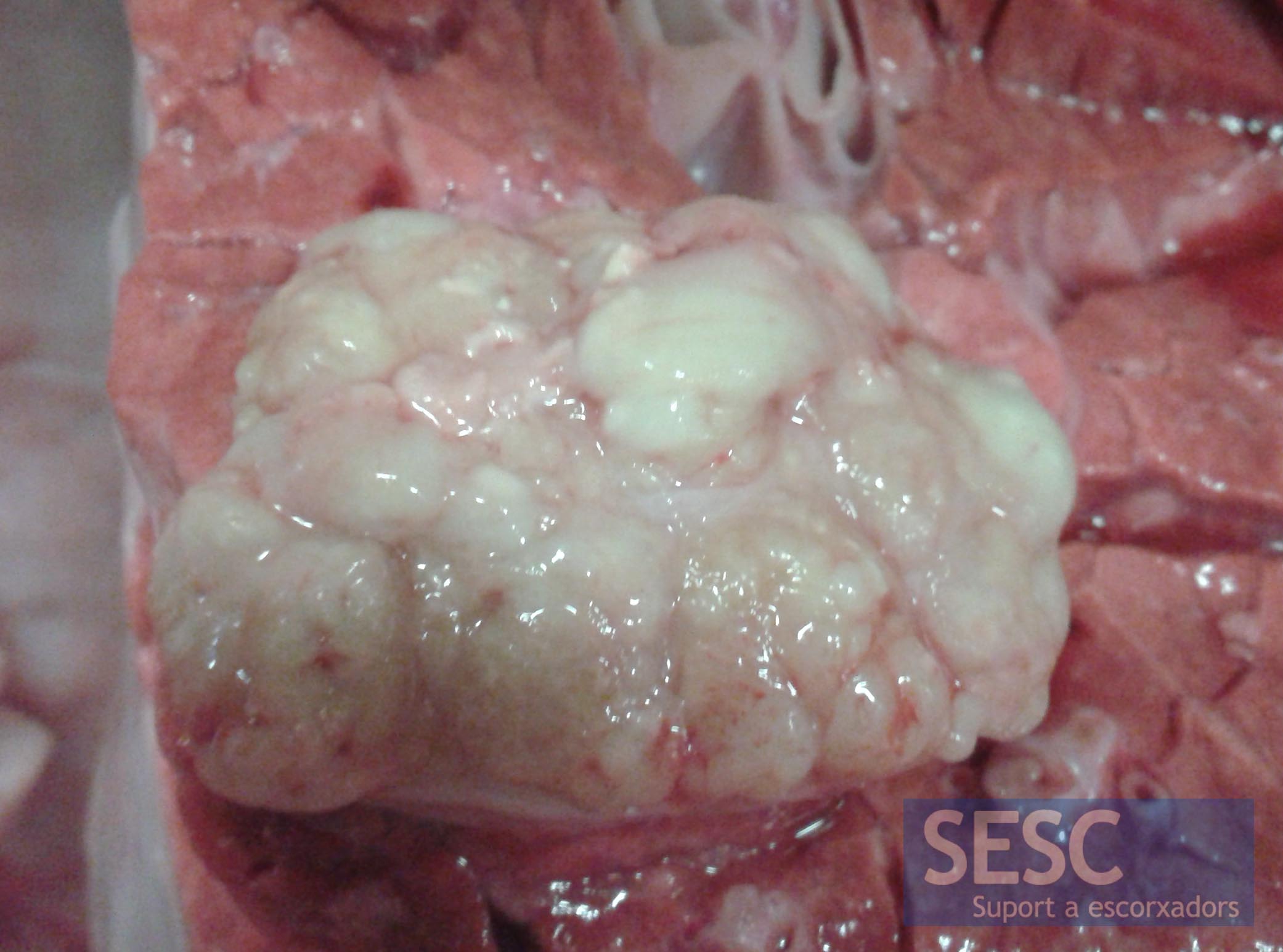
Detail of the lesion, which had a caseous appearance.
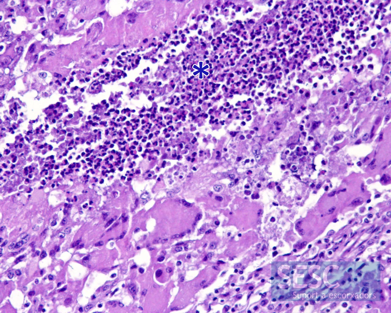
Histologically an intense suppurative infiltrate could be observed (asterisk) but also the presence of abundant macrophages, some them multinucleated (lower half of the image)..
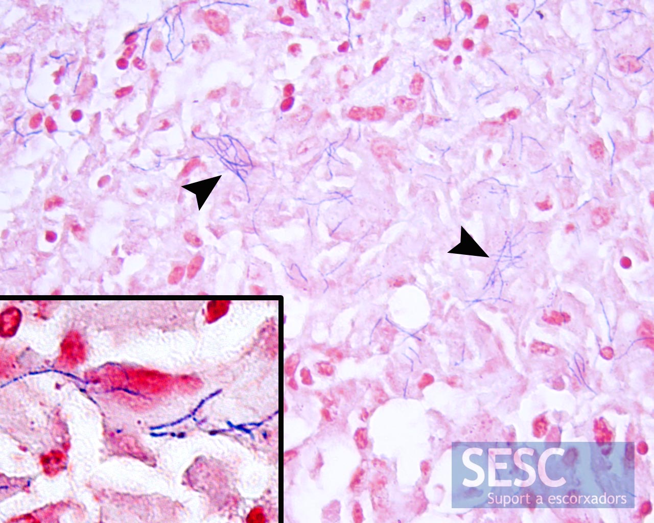
Gram staining allowed the observation of abundant gram positive filamentous bacteria (arrows), this can be seen in more detail in the insert on the bottom-left corner of the image.

Growth of Nocardia sp. colonies in blood agar from the lung sample.Image provided by the Servicio de Bacteriología y Micología del Departamento de Sanidad y Anatomía Animales de la UAB.
