What is your diagnosis? (19)
We present on this occasion, images of two cases (1 and 2) of granulomatous lymphadenitis in cattle.
Also, you can see images (A and B) of the corresponding histopathological studies.
Finally, you need to relate each image with its histopathology and the diagnosis you believe is correct.
Good luck!
CASE 1:
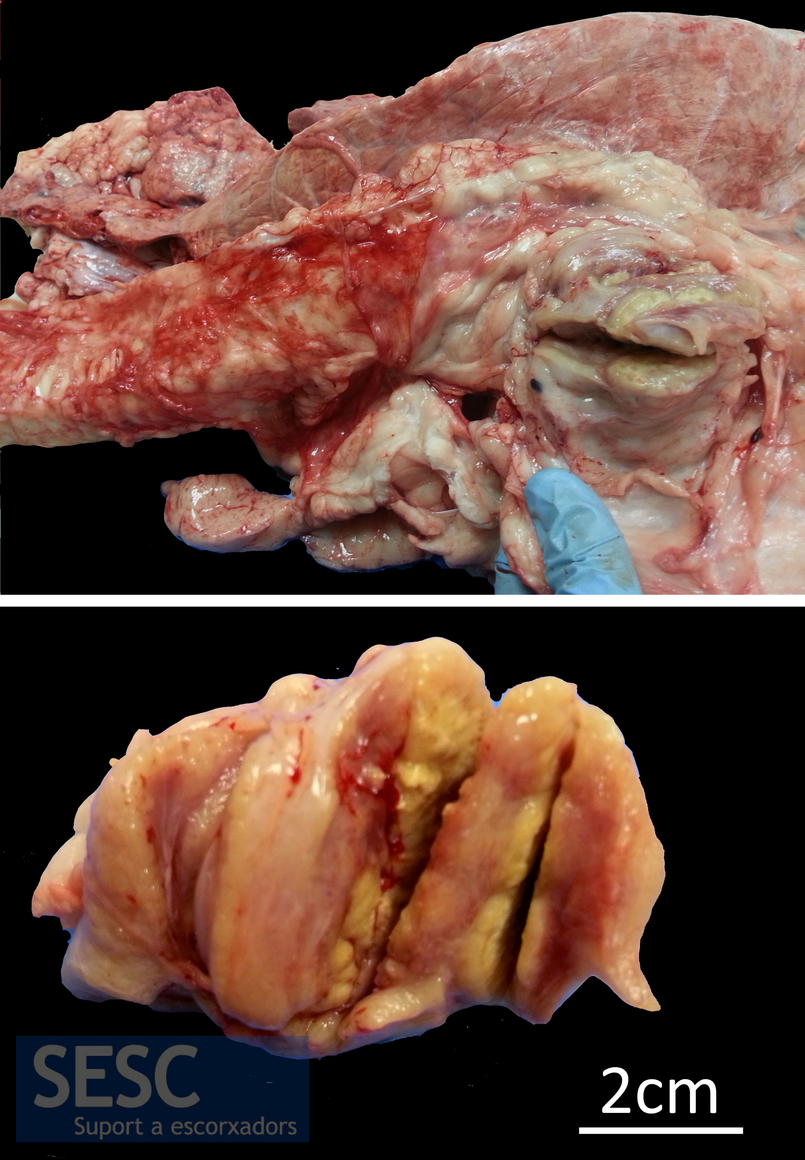
CASE 1: Granulomatous lesions in the traqueo-bronchial lymph node of a 13 months old, male, cross breed calf. In the picture below you can see the sectioned lesion.
CASE 2:

CASE 2: Granulomatous lesions in the lung parenchyma and mediastinal and retropharingeal lymph nodes in a 10 months old, male, Friesian calf.
HISTOPATHOLOGICAL STUDY A:
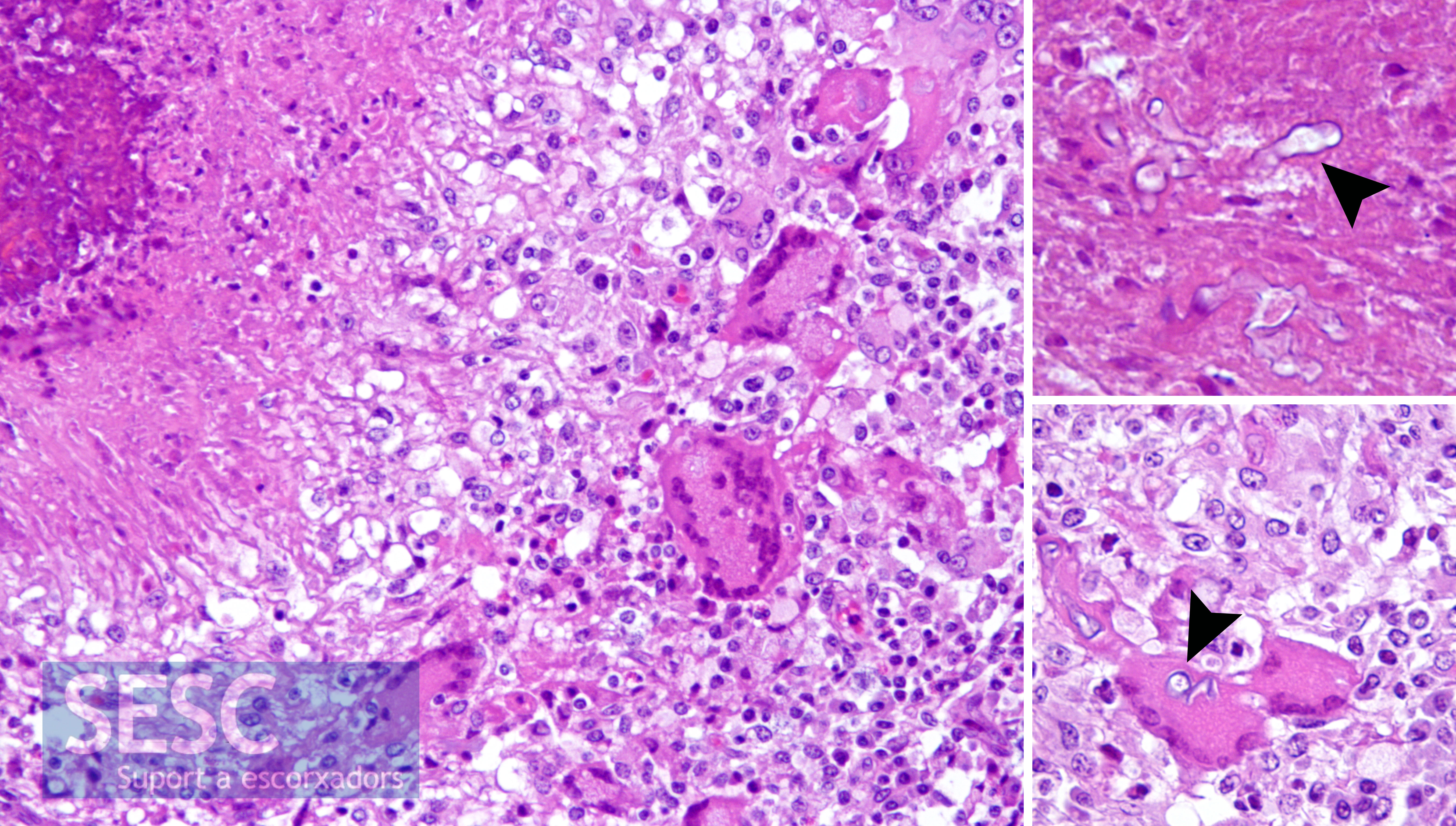
Histopathological study A: Presence of granulomatous inflammation with areas of necrosis, multinucleated giant cells and the structures pointed by the arrowheads (both in the necrosis and inside cells).
HISTOPATHOLOGICAL STUDY B:

Histopathological study B: presence of granulomatous inflammation with areas of necrosis, multinucleated giant cells and the structures that the arrowhead points at.