Jaundice in a sow
Histologically different liver lesional patterns were observed: areas of centrilobular necrosis with intense congestion of liver sinusoids (which correspond to reddish areas) alternating with irregular areas of coagulative necrosis with intralesional presence of bacteria (whitish areas). A discreet suppurative infiltrate was also present. The lesion thus corresponded to necrotizing hepatitis.
The massive release of hepatic origined bilirubin to the bloodstream, due to lesions described, explains the yellow discolouration of the carcass.
Gram staining disclosed gram negative and gram positive bacili. The presence of gas bubbles (from the products of anaerobic fermenttion) and the result of Gram staining indicates a bacterial cause, probably of the genus Clostridium, although a mixed infection cannot be excluded.
Unfortunately only formaldehyde fixed samples were submitted and the etiology could not be confirmed through microbiological culture.
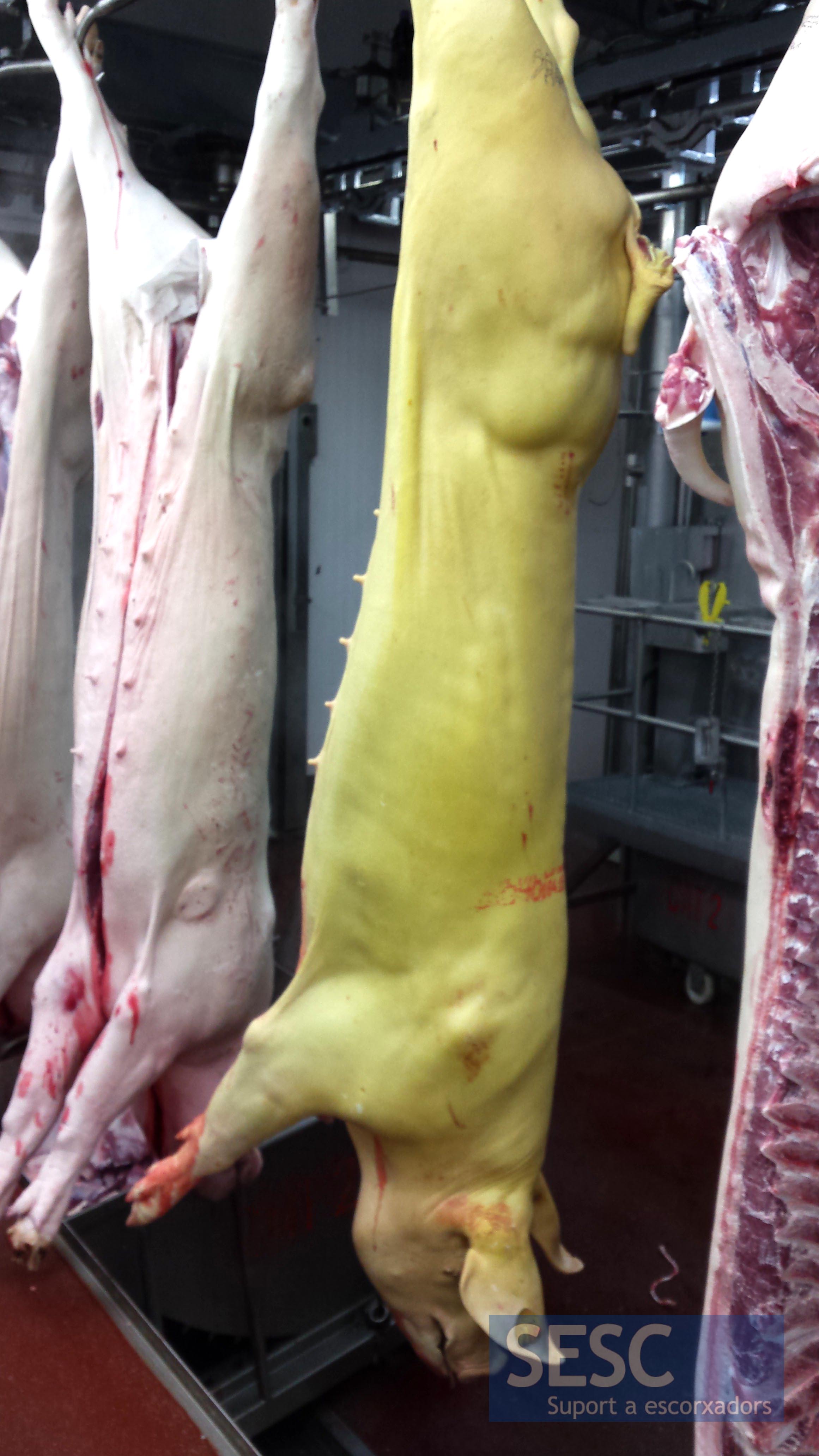
Generalised jaundice.
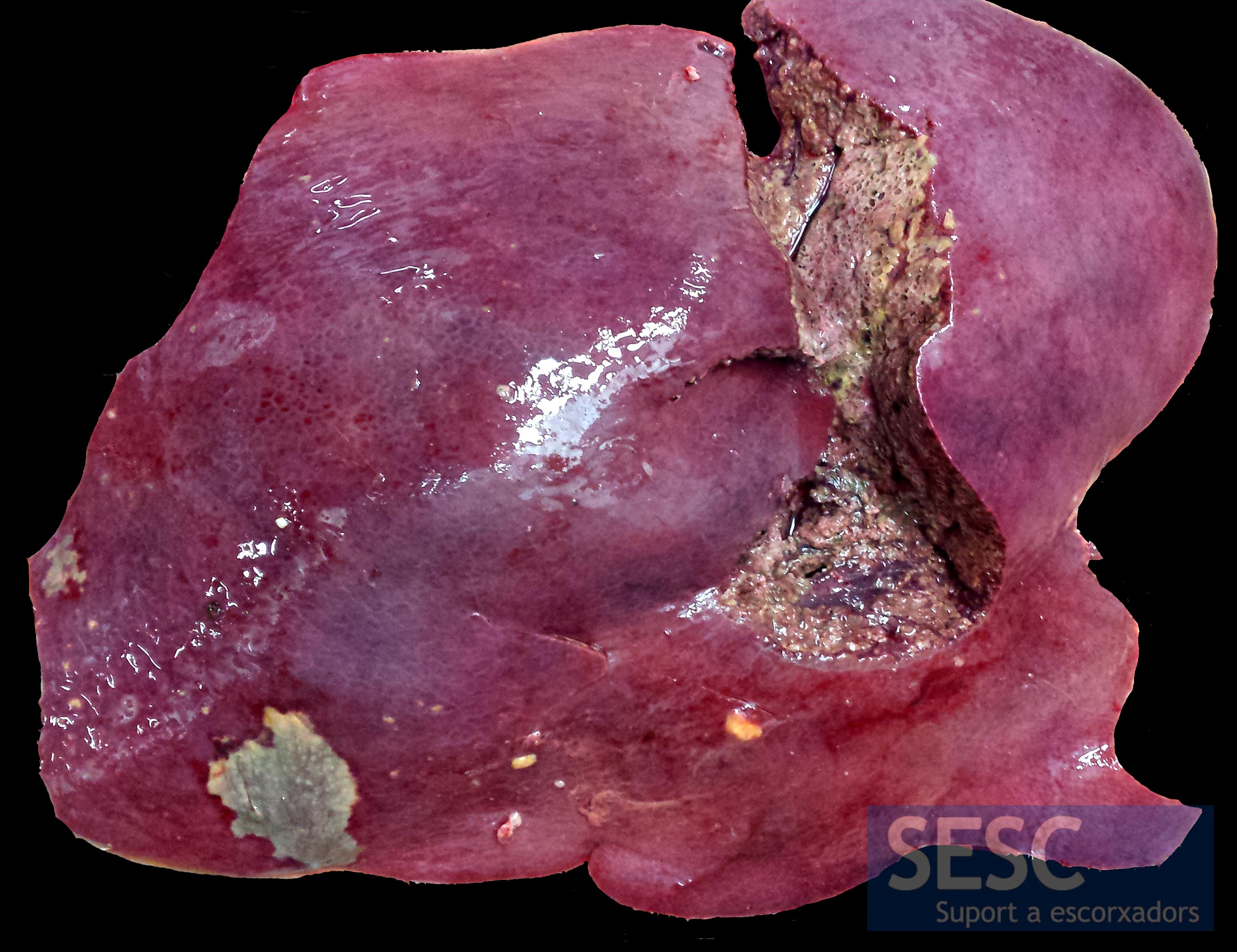
Liver aspect, friable to manipulation.

When sectioned gas bubbles and discoloration of the liver parenchyma were observed.

Detail of the lesion in the liver parenchyma (once fixed in formalin): reddish areas, white areas and gas bubbles.
