Mucormycosis in a calf
In a male, friesian, eleven months old calf two nodular lesions were observed, with a granulomatous appearance in the lung and caudal mediastinal lymph node.
Immediately, inspectors submitted samples to SESC to rule out Tuberculosis because the lesions are compatible with a Tuberculosis primary complex.
Histology confirmed that it was granulomatous lesions but showed the presence of non septate, irregular fungal hyphae within the lesions, some of them inside multinucleated giant cells. Groccot staining confirmed this finding.
It was not possible to isolate any fungus in microbiological culture, but a subsequent DNA extraction was performed and amplification and sequencing with universal 28S rRNA gene primers. The analysis of the sequence obtained compared with the GenBank database (BLAST) gave a 100% match with Rhizomucor pusillus (formerly known as Mucor pusillus), a mucoral (a fungus of the Order Mucorales).
A Mucormycosis (formerly known as zygomicosis) is a rare disease, it is not a contagious one, mucorales are found in the environment and can enter through respiratory airways (also digestive or through wounds in the skin). They usually affect immunocompromised individuals. Cases have also been reported in people. R.pusillus is thermophilic and thus grow in high temperature environments.
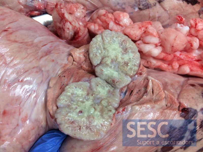
Granulomatous lesion in the lung parenchyma.
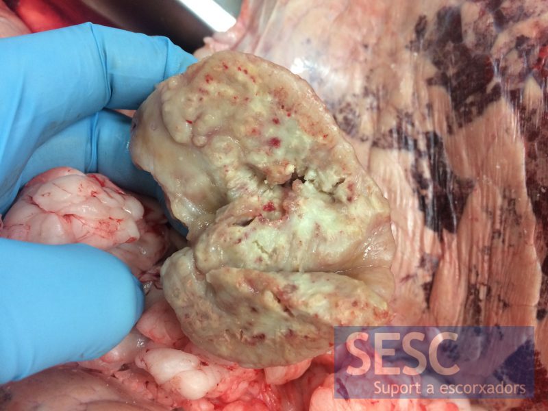
A similar lesion was observed in the caudal mediastinal lymph node.
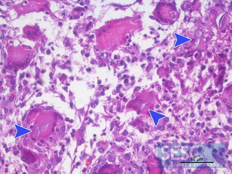
Histopathology shows granulomatous inflammatory infiltrate with structures compatible with fungal hyphae (arrowheads) some of them within multinucleated giant cells.
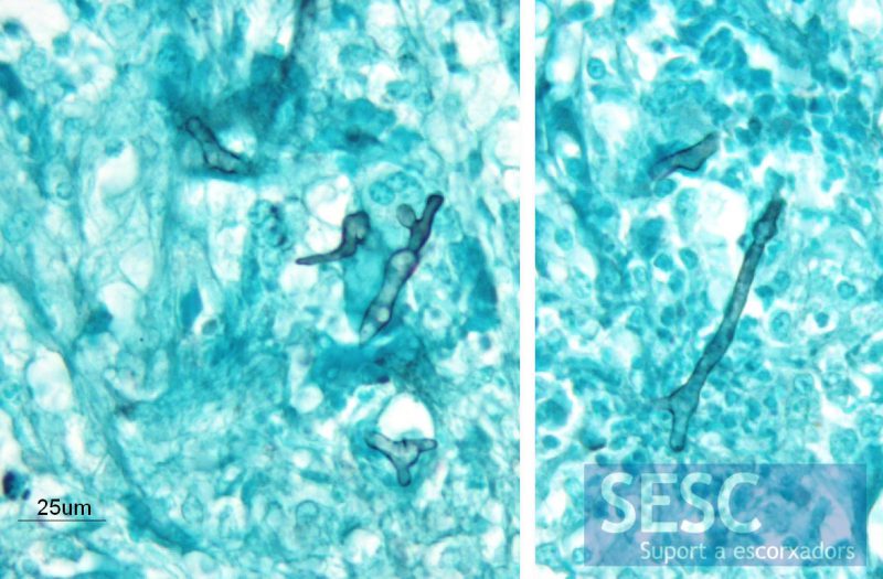
Groccott's stain confirmed the presence of irregular non septate hyphae (stained in black).
