Malignant peripheral nerve sheath tumour in a Friesian cow
Samples were submitted from a 3.5-year-old female Friesian breed cow carcass that presented multiple nodular lesions in the lung. The official meat inspectors identified the lesions as potential granulomas, prompting them to send samples to confirm their nature and rule out tuberculosis.
The nodular lesions were prominent, measuring between 0.5 and 1 cm in diameter, and were distributed throughout the entire lung parenchyma. Upon sectioning, they exhibited a firm consistency and a homogeneous whitish appearance, with no evidence of mineralized centers.
Histology was performed on some of the larger nodules, revealing a densely cellular, well-defined, and finely encapsulated neoplastic proliferation. This proliferation consisted of spindle cells arranged in fascicles and occasionally in bundles. Immunohistochemical analysis against chromogranin, a marker for neuroendocrine cells, resulted negative. However, positive immunohistochemistry against S100 protein (a marker for peripheral nerves) allowed the classification of the neoplasm as a malignant peripheral nerve sheath tumor .
In cattle, peripheral nerve sheath tumors are typically reported as incidental findings at the slaughterhouse, as they usually do not have clinical consequences for the animal. Nevertheless, the most common locations for this type of tumor are the nerves of the brachial plexus, heart, and intercostal and mediastinal nerves. While the lung contains nerves following the bronchial tree, they are in small quantities, and peripheral sheath neoplasms in the lung are extremely rare. Post-mortem examination did not identify any masses outside the lung; however, we cannot exclude the possibility that these are multiple metastases from a malignant neoplasm in one of its more common presentations. (ML, AC)
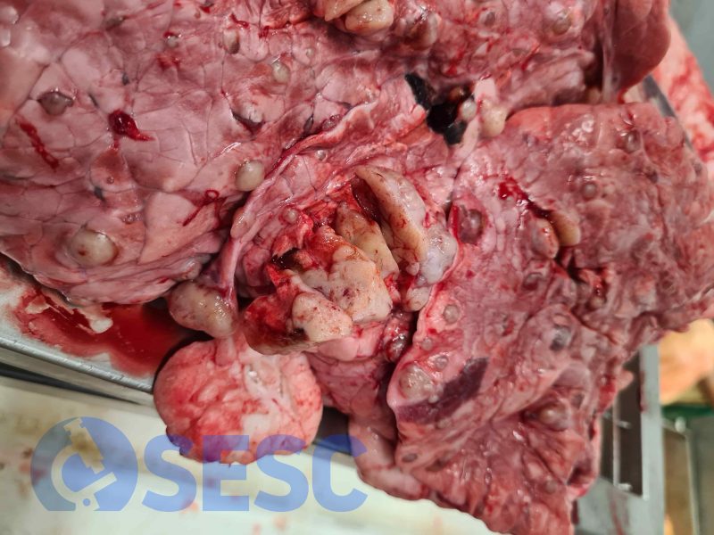
Cattle lung presenting a mass of 2-3 cm in diameter, as well as multiple smaller masses (approximately 0.5 cm in diameter) protruding over the pleura. When sectioned, they exhibited a homogeneous whitish appearance and firm consistency.
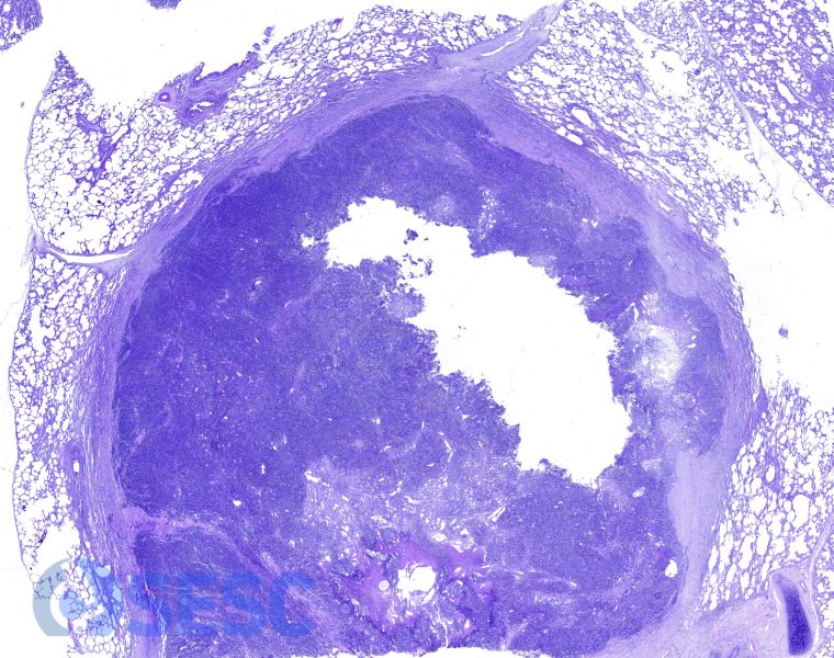
Histological image of one of the masses at low magnification. A densely cellular neoplasm was observed, well-demarcated from the adjacent lung tissue by a capsule of fibrous tissue.
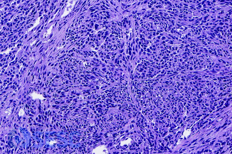
At higher magnifications, fascicles and bundles of small neoplastic cells were observed, with a nucleus showing condensed chromatin.
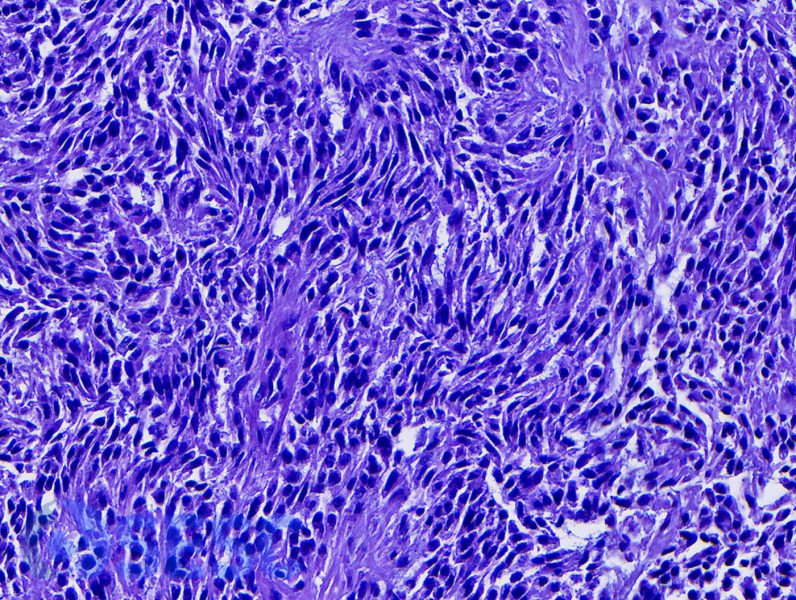
Occasionally, the nuclei of neoplastic cells were elongated and exhibited a palisading arrangement.
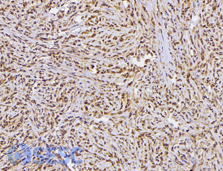
Immunohistochemical staining of the neoplasm against S100, revealed that the majority of neoplastic cells were positive for this marker. This result is indicative of its origin in peripheral nervous system cells.

1 comment(s)
Tumor maligno de origen genético