Chronic peritonitis in calves
These lesions correspond to a fibrous or fibro-adhesive peritonitis. This is clearly a chronic lesion. Probably a result of an old fibrinous peritonitis. It may be the sequel to poliserositis due to a systemic neonatal bacteremia (colibacillosis or streptococcus) or infection of the umbilical cord (the lesions in these cases are more severe on the ventral part).
These lesions may also occur as a result of viscera perforation such as the abomasum, the intestines, the reticle (traumatic reticuloperitonitis) or the uterus. Chronic peritonitis can also present with abdominal exudate and presence of suppurative abscessing lesions.

Plaques of fibrous material attached to the peritoneal serous membrane.
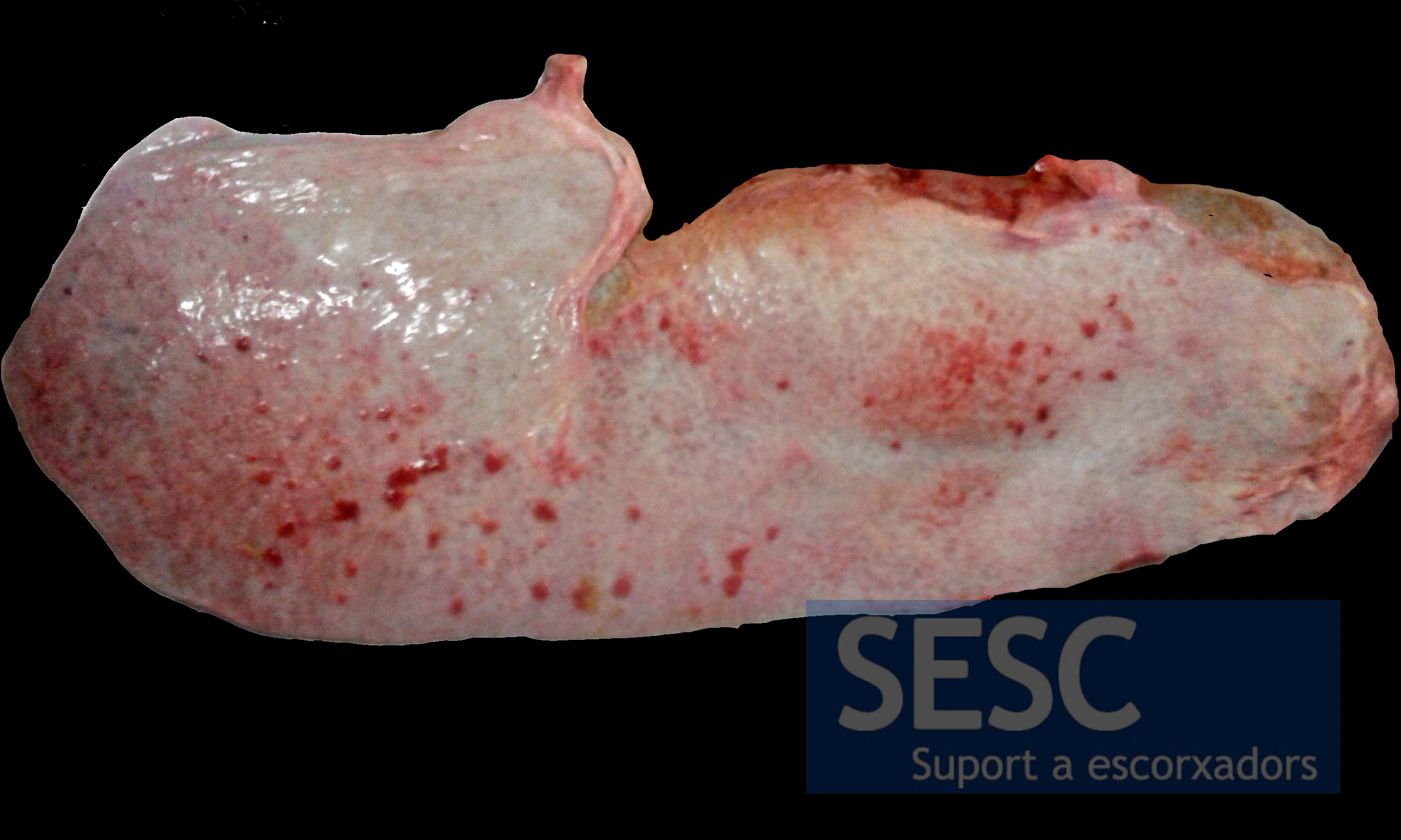
The spleen's serous membrane was also involved with multiple punctate reddish lesions.
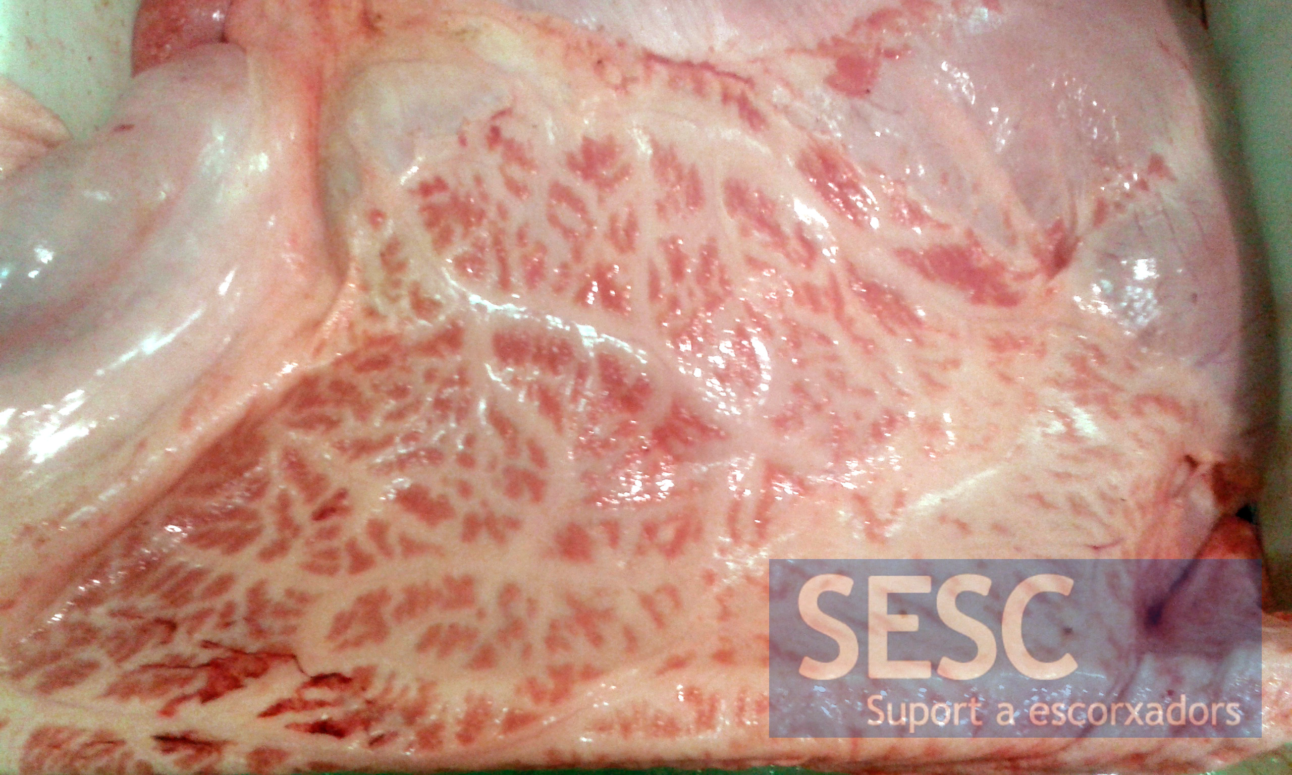
The mesentery also had deposits of fibrous material of reddish-pink coloration.

2 comment(s)
Para citar una página web sin autores puedes nombrar el nombre de la web y el año de publicación del post –> (SESC.cat, 2015).
En la lista de referencias hay que mencionar “Título del artículo.” Titulo de la web. Fechas de acceso (MM/DD/YYYY). URL –> “Peritonitis crónica en terneros”. SESC.cat. 08-25-2023, http://www.sesc.cat/es/peritonitis-cronica-en-terneros/
Revisa las particularidades de cada publicación pues puede variar el formato.
excelente información, me gustaria que se publicaran los autores de la información para poder citar