

27/07/2026
|
Bovine
0
Soft tissue sarcoma in the heart of a cow
Samples and images were submitted of the heart of a 10 years old, female cow, of the Alpine brown breed, in which, when performing sections on the myocardium to look for lesions of bovine cysticercosis, a whitish, sub-epicardial, focal, nodular lesion, between 0.5 and 1 cm in diameter was detected. The inspector wanted to rule out that it was bovine cysticercosis.
Microscopically, a neoplastic proliferation of spindle cells, well differentiated with the presence of fibrovascular stroma was observed. The lesion was classified as a low-grade soft tissue sarcoma, a neoplasm of connective origin. The suspicion of cysticercosis was ruled out.
Bovine cysticercosis is usually presented as cysts in the muscle tissue with an invaginated scolex inside or in the form of small granulomas in the muscles, both skeletal and myocardial, which macroscopically we see as whitish lesions. Within the differential diagnosis, we have described other processes such as: a myocardial infarction, a myocardial adenomatoid tumor, peripheral nerve sheath tumors, arteriosclerosis, benign mesenchymal proliferations similar to the one in this case, abscesses, lipomatosis cordis, lipoma, etc. (EV)
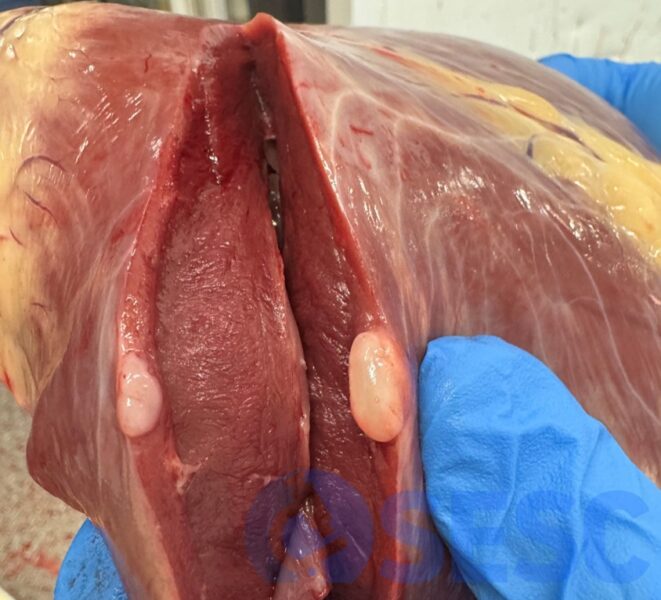
Whitish nodular lesion in the myocardium of a cow.
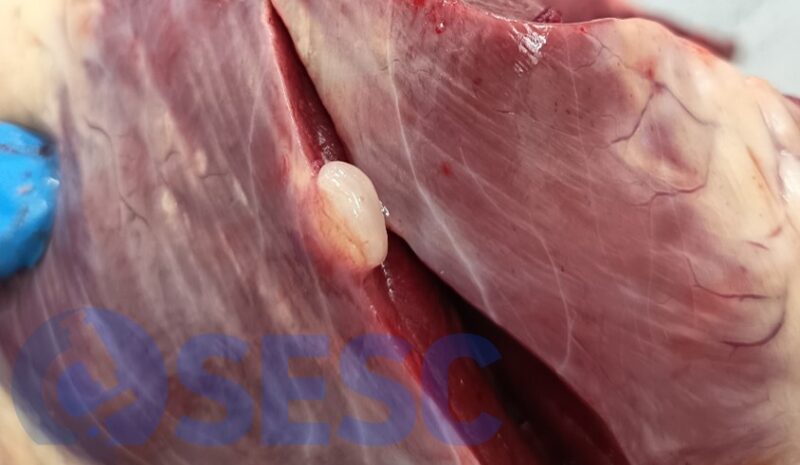
Detail of the sub-epicardial lesion.
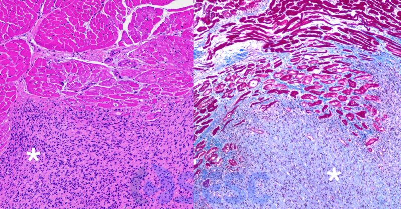
Microscopic image of the lesion. On the left, hematoxylin and eosin staining shows the fusiform neoplastic proliferation (asterisk) that infiltrates the muscle fibers seen at the top. On the right, Masson's trichrome staining shows the collagen-rich stroma (blue) of the proliferation.