

Myeloma in a bovine carcass
We received a case of a bovine carcass (Asturian breed) aged 14 months, presenting a generally altered appearance. With a diffuse distribution, the musculature and subcutaneous tissue exhibited marked congestion, while the lymph nodes showed blood resorption. The viscera also displayed significant congestion, and petechiae were observed in the kidneys. Upon longitudinal sectioning of the carcass, a notable loss of consistency was observed in the central part of the medullary cavities of the sternebrae and vertebrae. Samples from various viscera were submitted to the SESC for further study and diagnosis.
After receiving the samples, decalcification and histological analysis were performed. The bone marrow, both from the sternebrae and vertebrae, exhibited a neoplasm composed of round cells, proliferating and occupying the medullary cavity, replacing nearly all the hematopoietic tissue and bone spicules. In other words, the neoplastic growth had destroyed the bone tissue, a process known as an osteolytic lesion.
An immunohistochemical study was conducted, demonstrating that the cells were positive for CD20 (a B-lymphocyte marker) and lambda (an immunoglobulin component used to identify plasma cells). Additionally, the cells were negative for CD3 (a T-lymphocyte marker). Based on these characteristics, a diagnosis of myeloma (a plasma cell neoplasm in the bone marrow) was established.
Although this neoplasm is extremely rare in bovines, its presentation is similar to that reported in other species where it is more common. In fact, the presence of osteolytic lesions in the medullary cavity of the vertebrae is the most characteristic macroscopic feature of multiple myeloma in dogs. (AC)

Carcass from a bovine. General appearance of the carcass, in comparison with normal appearance a carcass (behind). A generalized congestion /reddening of the musculature and the adipose tissue can be observed.
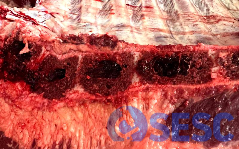
Carcass from a bovine. When the carcass is sectioned in halves, the sternebrae show a osteolytic lesion where the normal tissue is replaced by a reddish friable mass.
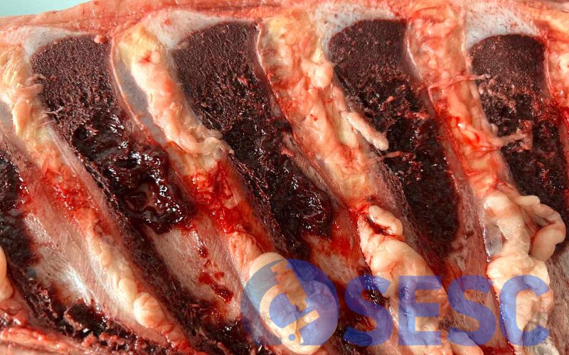
Carcass from a bovine. The same osteolytic lesion, with marked loss of consistency, can be observed in the bone marrow of the spinous process.
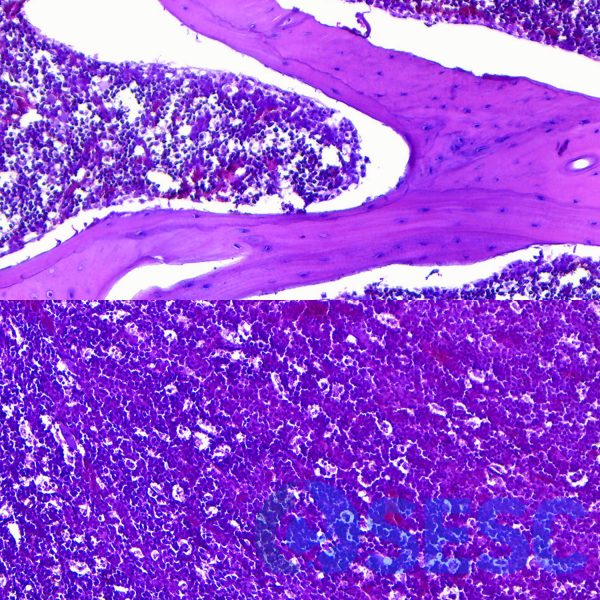
Histologic section of the lesion at the vertebral body. The top picture shows the normal tissue at the periphery of the vertebral body, displaying bone trabecula forming medullary cavities, where the hematopoietic tissue is found. The image at the bottom consists of the centre of the vertebral body, showing replacement of the bone tissue by a proliferation of round cells, consistent with plasma cells, which explains the macroscopic changes observed.