Nodular lesions in the subcutaneous tissue of a bovine carcass
Images were received of a bovine carcass presenting nodular lesions with a granulomatous appearance in multiple areas of the subcutaneous adipose tissue. The lesions did not extend into the underlying musculature. Their distribution was generalized, involving the neck, dorsal region, and hindquarter of the carcass. The dermis and epidermis were not affected. The lesions were well demarcated and ranged from 0.5 to 2 cm in diameter.
Histologically, the lesions corresponded to foci of necrotic tissue located within the adipose panniculus, associated with areas of mineralization, fibrosis, and a moderate mononuclear, angiocentric inflammatory infiltrate. These lesions were nonspecific and were not associated with any identifiable etiological agent. However, in situ hybridization confirmed the presence of small amounts of RNA from the lumpy skin disease virus (LSDV).
As lumpy skin disease (LSD) should be included in the differential diagnosis of generalized nodular skin lesions, even when confined to the subcutaneous tissue, samples were submitted in parallel for PCR testing. These included EDTA blood, ocular, nasal and oral swabs, as well as samples from the nodular lesions. The results for each sample type are shown in the table below and demonstrate that the lowest Ct value (i.e., the highest viral DNA load) was detected in the nodule sample. It should be remembered that this is consistently the sample of choice for the diagnosis of LSD. The remaining test results indicated that the animal was not viraemic but was still shedding the virus.
It is noteworthy that the animal originated from an LSD-vaccinated herd. Therefore, a DIVA PCR (Differentiating Infected from Vaccinated Animals) assay was performed, as the attenuated vaccine strain can, in a very small proportion of cases, induce lesions similar to those caused by the disease itself. In this case, the analysis confirmed that a field strain was responsible for the lesions. The chronic nature of the lesions suggests that the animal was most likely vaccinated after it had already become infected. This practice should be avoided, as the PCR results demonstrate that vaccination under these circumstances does not prevent continued viral shedding. Consequently, if such an animal comes into contact with unvaccinated animals, it could give rise to a new outbreak. (EV)
| Sample | Capripoxvirus genus PCR | DIVA PCR |
| EDTA-blood | Negative | Not done |
| Ocular swab | Inconclusive (Ct=36,4) | Positive to field strain |
| Nasal swab | Positive (Ct=33.6) | Positive to field strain |
| Oral swab | Negative | Not done |
| Subcutaneous lesion | Positive(Ct= 13,2) | Positive to field strain |
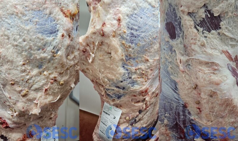
Bovine carcass with multifocal nodular lesions in the subcutaneous tissue, firm to the touch and showing a necrotic appearance.
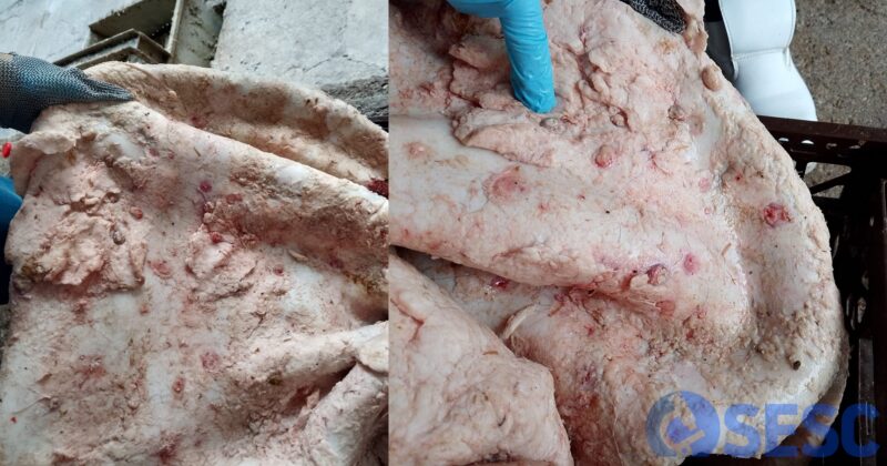
Appearance of the lesions attached to the hide.
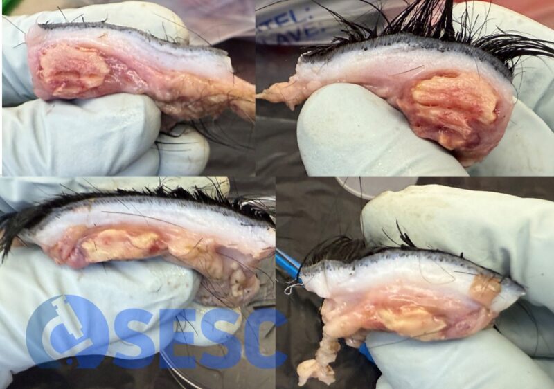
Cross-sectional appearance of the nodules. The lesion was confined to the subcutaneous tissue and showed a necrotic appearance. No other lesions were observed in the internal organs.
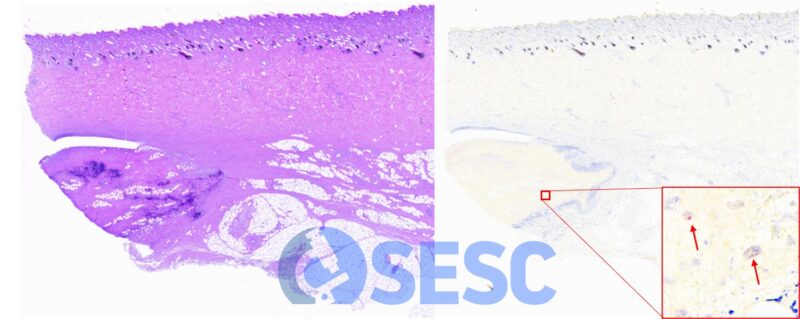
Histological appearance of the lesions, corresponding to areas of necrotic tissue with mineralization (dark blue staining), fibrosis, and remnants of a mononuclear inflammatory infiltrate. The image on the right shows the results of in situ hybridization, demonstrating the presence of intracellular cytoplasmic viral RNA (arrows) in very small amounts within cells of the inflammatory infiltrate.