03/07/2012
|
Liver
0
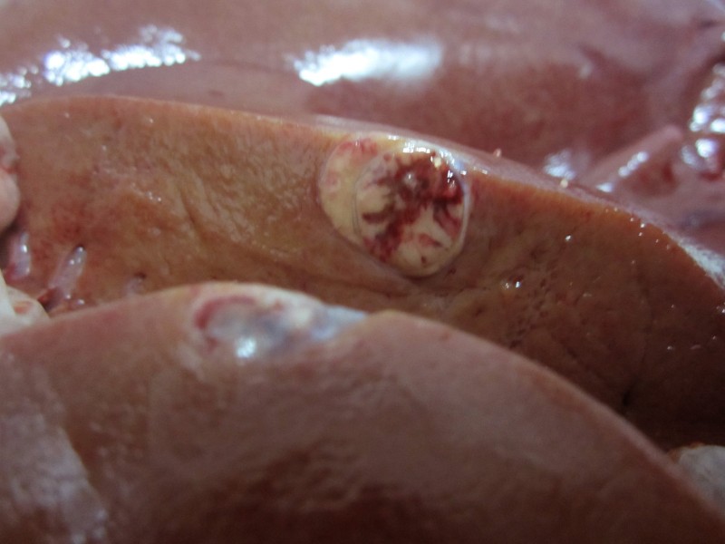
Intraparenquimatous hepatic nodule
The cut surface was pale with haemorragic areas. The lesion was encapsulated and well circumscribed.
Due to its intraprenquimatuos location the meat inspectors main suspicion was of a a hydatid cyst altered by the host inflammatory reaction.
The histopathological study revealed it was a benign neoplastic proliferation of epithelial origin, probably derived from hepatocytes, therefore a diagnosis of hepatocellular adenoma or hepatoma was established.


