Chronic vascular lesions in a pig kidney
We received an inquiry regarding a 6 month-old, hybrid breed, swine carcass that showed multifocal whitish areas lesions in both kidneys. Samples from both kidneys and renal lymph nodes were submitted to the SESC for their diagnosis, in which a chronic interstitial nephritis was initially considered due to their macroscopic appearance.
When the samples were received, the appearance of the lesions indeed consisted of multifocal white areas and rugosity/irregularity of the renal surface. However, the distribution and shape of the lesions was peculiar, as they were preferentially centred around the corticomedullary junction and displayed lineal shape.
When the histopathological study of the samples was performed, the lesions did not correspond to inflammatory infiltrates within the renal interstitium as initially considered. They consisted of small and medium calibre arteries that were markedly thickened and distorted. They displayed a wide range of degenerative/proliferative alterations, and to a lesser extent, inflammation, which included hyperplasia of all tunica, vacuolization of smooth muscle cells, obliteration of the vascular lumen with recanalization phenomena, fibrosis of the adventitial layer with occasional mononuclear inflammation, and hyperplasia of the vasa vasorum. This constellation of vascular alterations is classified as a diagnosis of severe generalized arteriosclerosis with mild periarteritis. Additionally, scant glomeruli displayed fibrinoid degeneration.
At the histologic study the lymph node did not present lesions within the lymphoid tissue itself, but the arteries present within the histologic section displayed the same alterations as those observed in the kidney. For this reason, it is highly probable that the observed alterations were systemic in nature and that they were present in other organs. However, this could not be confirmed as other viscera were not submitted.
Regarding the etiopathology of this lesions, it must be noted that they are not frequent lesions in swine, and that they consist of a chronification of a intense vascular damage centred in muscular arteries, which most probably was necrotizing and/or proliferative in nature. In other words, we observed a scarring of old vascular lesions. For this reason, the most probable explanation is that this consisted in the chronification of a porcine dermatitis and nephropathy atypical case (with almost absence of glomerular alteration). (AC)
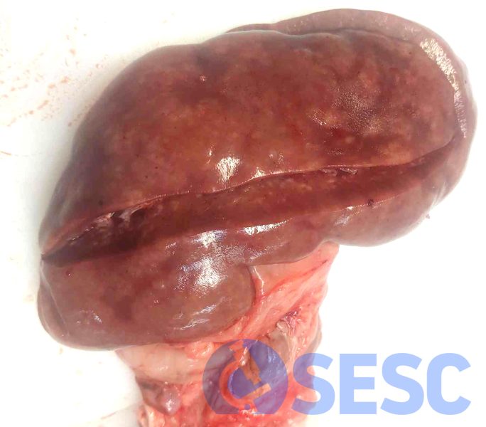
Kidney from a pig. An irregular and rugous surface with multifocal white areas can be observed.
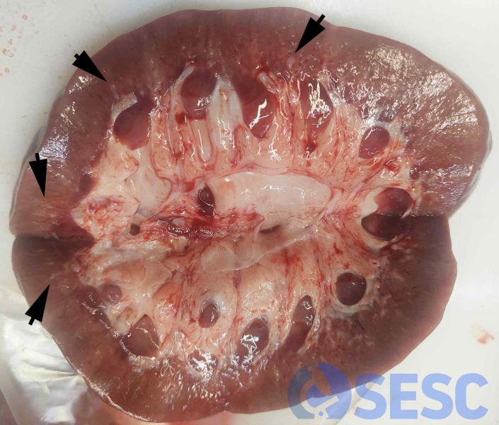
Kidney from a pig. At cut section, multiple white lesions can be observed mostly centred around the corticomedullary junction, often presenting linear morphology (arrows).
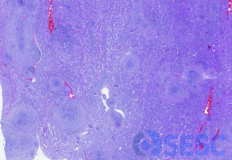
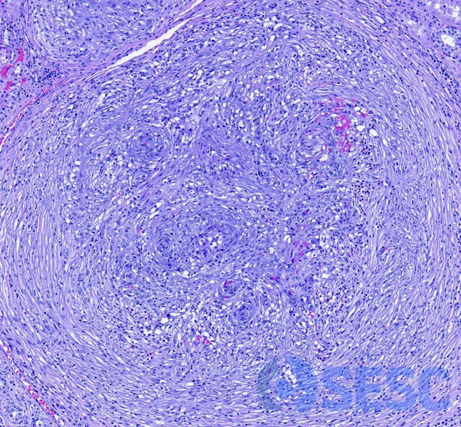
Histologic picture at low magnification, which showed that the white areas corresponded to vascular alterations (affecting muscular arteries of low and medium calibre) which markedly increased their size.
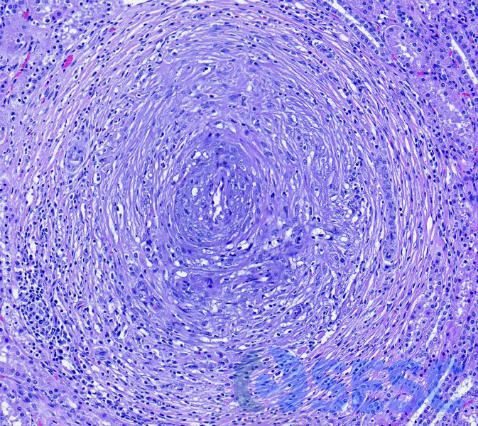
One of the arteries, in which it could be observed a marked expansion of all the tunica (hyperplasia-hypertrophy), with proliferation of vasa vasorum, vacuolization of the smooth muscle cells, fibrosis of the adventitial layer and mononuclear infiltrates.
Artery which showed similar changes as those mentioned earlier, however in this case the central lumen was substituted by different vascular canaliculi of lesser size. This is indicative of arterial recanalization, i.e. lumen neoformation posterior to a thrombosis.
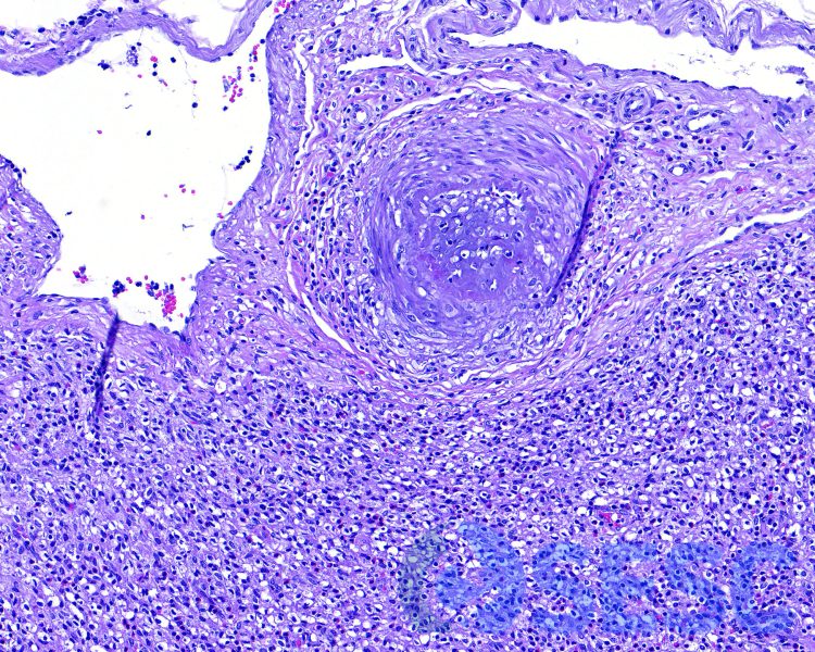
Similar lesion in a muscular artery in the capsule of the lymph node. Although other viscera were not studied, the appearance of these lesions in both the kidneys and lymph nodes suggest a systemic disease.
