

Necrosis of the lumbar axial musculature in a pig carcass
We received an inquiry regarding a 6-month-old hybrid breed pig carcass, where the lumbar axial musculature exhibited a yellowish coloration and a fibrous appearance. Samples of the axial musculature, spinal cord, lymph nodes, and vertebral bone were submitted.
Upon receiving the samples, we observed that the skeletal musculature had a focal, extensive area of yellowish discoloration and hardened consistency. The rest of the tissues did not show any obvious macroscopic lesions. In the histopathological study, it was observed that the affected area corresponded to muscle necrosis with loss and replacement of muscle fibers with fibrous tissue. Among this proliferation, there were remnants of degenerated and occasionally mineralized muscle fibers, surrounded by large satellite cells with abundant nuclei. In the fibrotic areas, there was also a marked proliferation of granulation tissue and the presence of lipid vacuoles of variable size.
The specific cause of the observed muscle lesion could not be determined; however, despite being an unusual location, it cannot be ruled out that it was a consequence of the chronic injection of irritating products in this area. The spinal cord, lymph nodes, and bone did not show any lesions, suggesting that this was an exclusively muscular process. (ML)

Lumbar axial musculature of the pig. A focal, extensive area of yellowish coloration was observed.

Fragment of the lumbar axial musculature of the pig. The affected area corresponds to the more dorsal half of the sample fragment.

Histological image at low magnification, showing hypereosinophilic, retracted, and some mineralized muscle fibers (darker purple coloration).

Fibrotic area showing marked proliferation of granulation tissue and the presence of lipid vacuoles of various sizes.
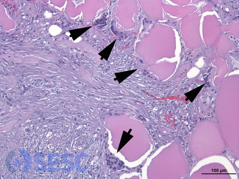
Numerous active satellite cells (arrows) were observed around the muscle fibers.