White spherical lesions in the abdominal viscera of a tuna
An enquiry was submitted by an inspector of a fishermen's guild that observed multiple spherical white lesions in a red tuna (Thunnus thynnus) liver.
Once the lesion was examined more closely, it was appreciated that in fact, the white lesions were mostly located in the serous membrane and between the connective tissue of the pyloric caeca. The squash extension of one of these spherical structures, observed under the microscope, allows the observation of a multitude of microsporidia spores of 3-4 microns in diameter that, morphologically, were compatible with descriptions of similar species (such as japenese seriola, Seriola quinqueradiate) of microsporida of the genus Microsporidium.
These parasites (of the fungi kingdom) are intracellular and cause hypertrophy of host cells due to the presence of their spores forming the macroscopic spherical lesions that are generically called xenomas.
Microsporidia in fish are not known to cause any zoonosis. However, in immunosuppressed patients, opportunistic infections of some species of microsporidia have been described.
To do this diagnosis, we have incorporated to the SESC team the experts of the Fish Pathology Diagnostic Service (SDPP) of the UAB's Veterinary Faculty.
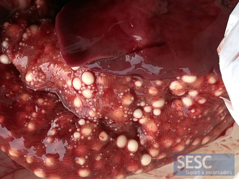
Multiple whitish spherical lesions (xenomas) to the pyloric caeca.
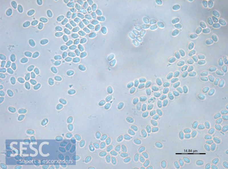
Squash extension of one of the xenomas, multitude of spores with the morphology and typical size of Microsporidia spores can be observed.
