Any given day in a ruminant slaughterhouse (2023)
This June we organized the yearly practical workshop on identification and description of lesions for official meat inspectors.Therefore, we collected viscera declared unfit for human consumption from two ruminant slaughterhouses, which were the subject of discussion during the session. In this post, some lesions that we were able to observe, and of which samples were also collected in formalin for histopathological study, are discussed. (AC)
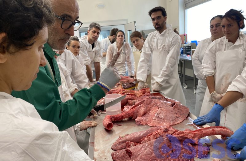
SESC collaborating pathologist Dr. Mariano Domingo (IRTA-CReSA, UAB) participated in the discussion of the lesions observed in viscera declared unfit.
Case 1
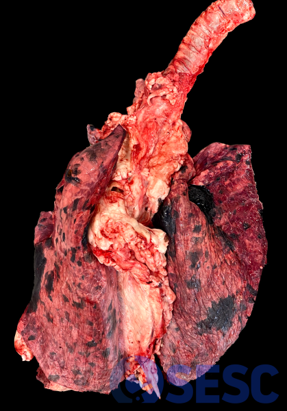
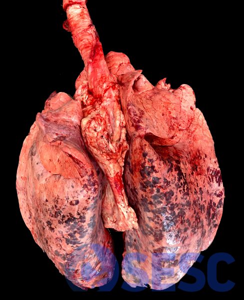
Bovine lung. Widespread multifocal areas of very dark staining can be seen in this bovine lung. On section, they go deep into the adjacent lung parenchyma.
Due to the distribution, they seem to affect entire lung lobules (lobulillar pattern). This dark coloration in fresh viscera (not fixed) is almost unequivocally due to melanin.
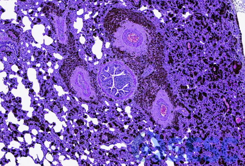
In the histological study, this can be easily confirmed, observing multiple aggregates of melanocytes with intracytoplasmic black pigment (melanin) in alveolar and peribronchial septa, etc.
This finding is common in slaughterhouses and is usually called macular melanosis, and corresponds to a defect in the migration of melanocytes, which end up locating in different viscera (lung, spleen, meninges...). It does not represent a dangerous alteration for the animal or for food safety, however, viscera are usually declared unfit for presenting alterations of the normal appearance.
Case 2
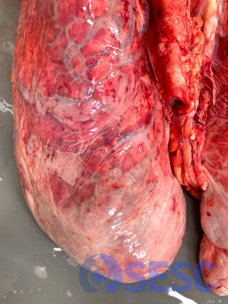
Bovine lungs. In this case, an alteration is observed that predominantly affects the diaphragmatic lobes, consisting of a marked distension of the pulmonary interlobulillar septa due to emphysema.
To distinguish it from emphysema that affects the alveoli (alveolar emphysema), this emphysema is called interstitial emphysema.
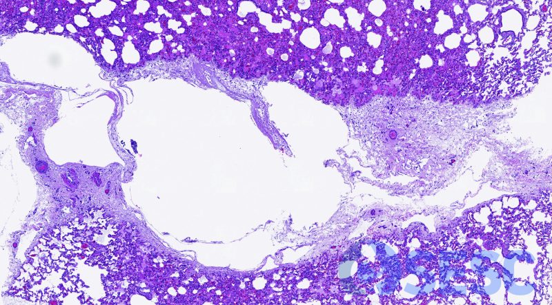
Histologically, it corresponds to the distension of the interstitial septa by air (without dye properties). This air dissects the interstitium of the septum, and dilates the lymphatic vessels present there.
Although interstitial emphysema can be a relevant finding in some conditions (bovine respiratory syncytial virus pneumonia; among others), the most common scenario at the slaughterhouse is that it is an incidental finding. However, the exact mechanism by which bovine lungs develop this emphysema in the slaughter process is not known.
Case 3
Again we find incidental post-mortem alterations in bovine lungs with little pathological relevance. In this case we see generalized reddened multifocal areas over the lung; much more pronounced on the dorsal edge of the diaphragmatic lungs.
In this case it consists of aspiration of blood during the sacrifice process. It should not be confused with aspiration pneumonia, since in this case an inflammatory component is absent.
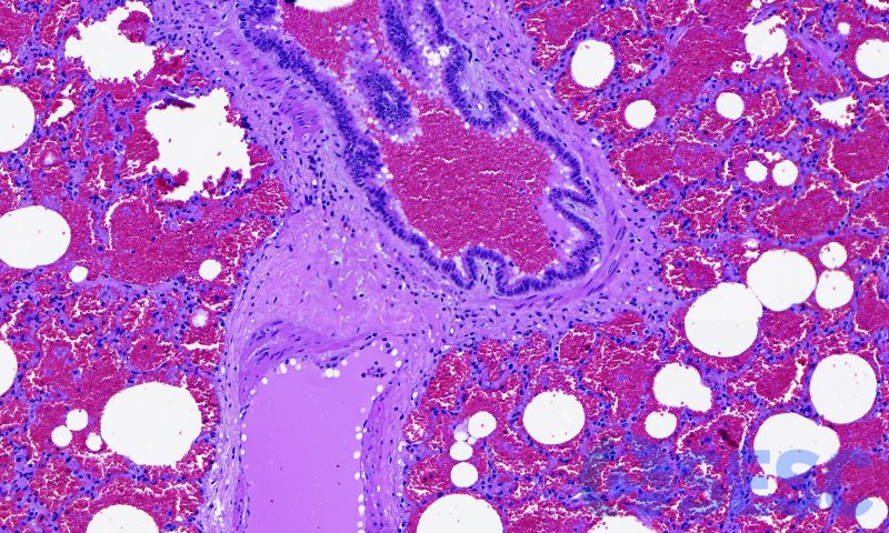
Histologically, we can confirm that this is blood aspiration because, in addition to finding abundant erythrocytes in the alveolar lumens, they also fill the bronchi and bronchioles of the affected lobule.
Case 4
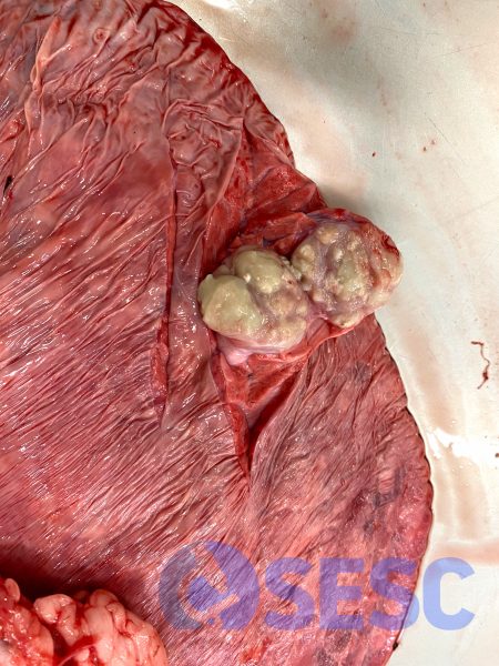
Bovine lungs. We found a focal lesion located in the pulmonary diaphragmatic lobe. It was hardened and prominent on the pleural surface, and when sectioned it slightly crackles, and presents multifocal areas filled with whitish purulent to caseous material.
This lesion is the prototype of a granulomatous lesion potentially compatible with tuberculosis, and it is important to study it in all cases (if found, samples must be sent to the laboratory). However, given the tuberculosis-free status of cattle herds in Catalonia; and the presence of a lung lesion in the absence of a lesion in the thoracic lymph nodes, this lesion is highly unlikely to be tuberculosis.
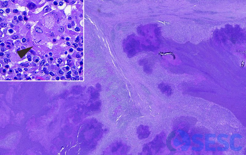
Histologically, the lung tissue can be observed almost completely replaced by irregular clusters of viable and necrotic neutrophils, delimited by abundant macrophages, multinucleated giant cells and granulation and fibrous tissue; which is accompanied by abundant mononuclear inflammation. In addition, in this focal pyogranulomatous pneumonia, bacilli can be seen in the cytoplasm of multinucleated giant cells, occasionally organized in a row (filamentous bacteria; probably Nocardia spp. arrow within inset). Therefore, this lesion did not correspond to tuberculosis.
Case 5
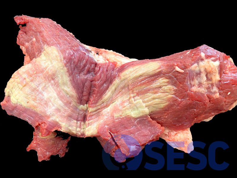
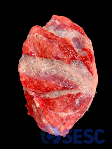
Bovine muscle In the section of the muscle (location not reported), multifocal coalescent striae of wide extension, whitish to slightly greenish in colour, are observed.
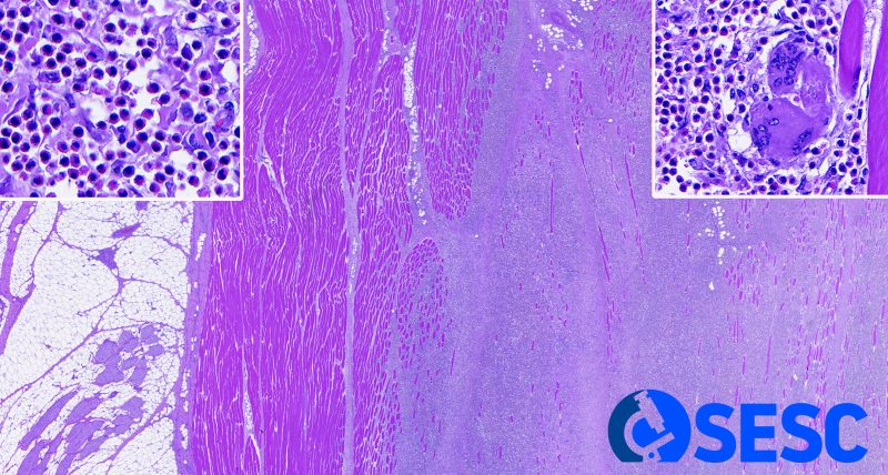
Histologically, they corresponded with loss of muscle fibers and replacement by abundant inflammatory infiltrate; as well as granulation tissue, fibrosis and neovascularization. The inflammation corresponded mainly to eosinophils (left inset), with abundant mononuclear cells and occasional multinucleated giant cells (right inset).
The histological findings are consistent with eosinophilic myositis or green muscle, which is an eosinophilic inflammation of bovine muscles. Although it is usually associated with Sarcocystis spp., an association with these parasites is not always observed, so the etiology has not been fully defined.

1 comment(s)
In fact regarding eosinophilic myositis, a study by De Bosschere and Ducatelle found and inverse relation between eosinophilic myositis and the number of Sarcocystis cystozoits in the heart tissue in cattle.