Chronic fibrino-hemorrhagic pericarditis in a calf
Histologically a fibrinous exudate was observed in reorganization, with formation of cubic epithelium coated pseudopapillae (reactive mesothelium). A moderate lymphoplasmocytic infiltrate was present along with macrophages and a few neutrophils. The lesion was classified as a chronic fibrinous pericarditis.
Although the type of lesion indicated a bacterial etiology, on microbiological culture no bacteria could be isolated associated with this type of pathology. This is most likely due to the chronicity of the lesion.
One possible explanation for this type of lesion would be a traumatic pericarditis (caused by the presence of a sharp object in the reticulum).
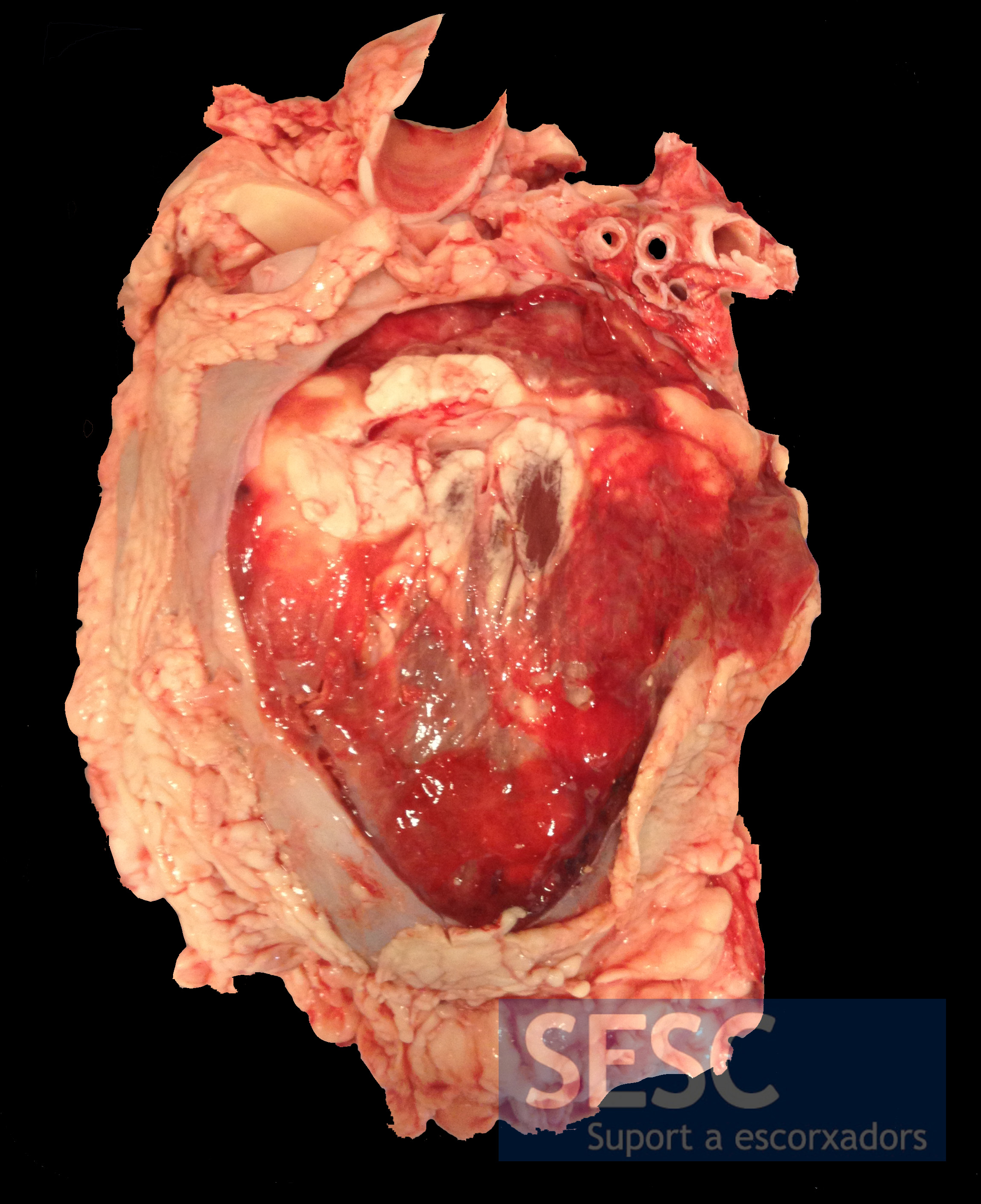
Fibrinous pericarditis. Picture of the sectioned Pericardium.
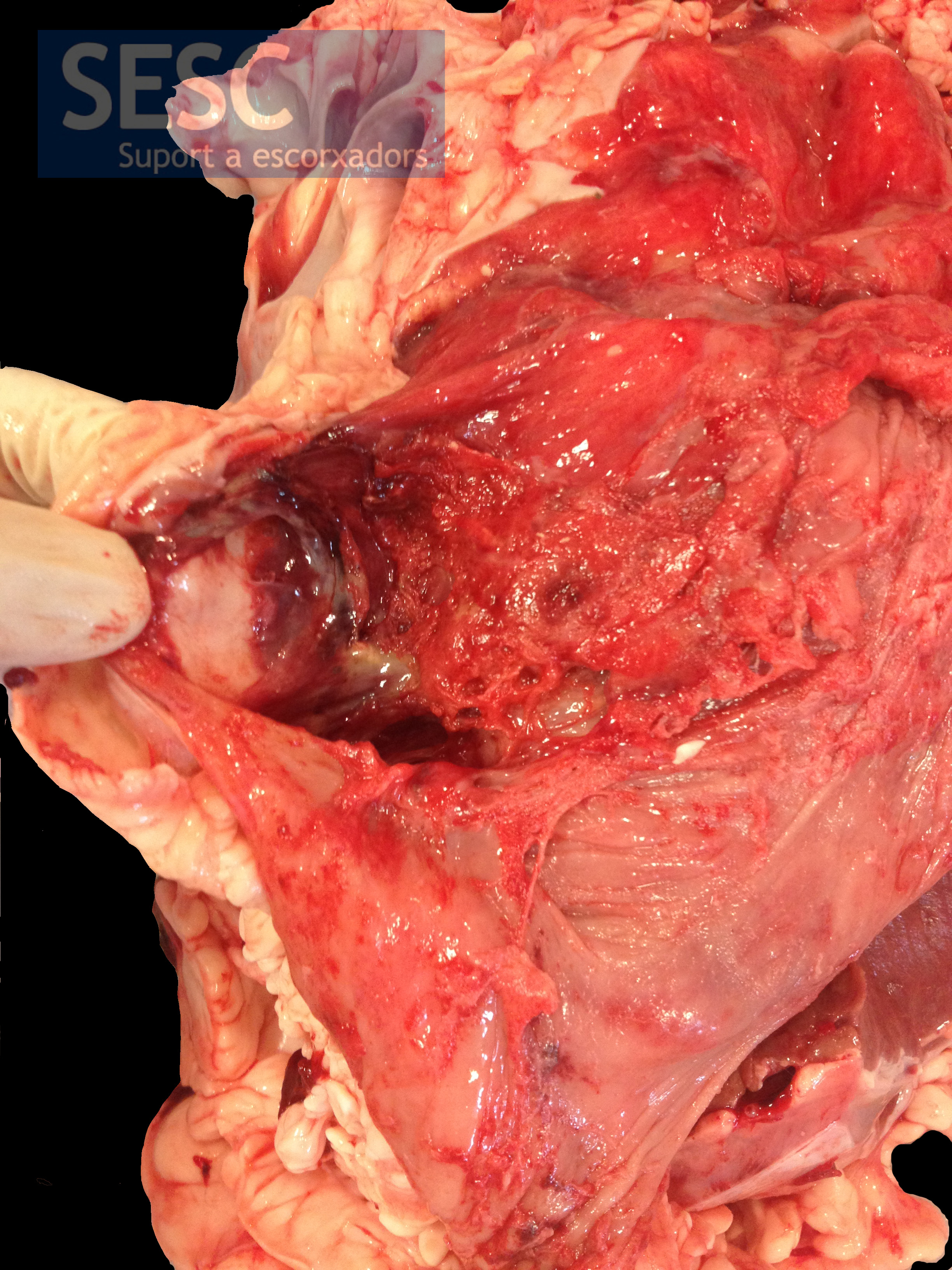
Detail of the inflammatory exudate.

2 comment(s)
Comment form Veterinary pathology group in LinkedIn:
Very nice specimen.If it is due to traumatic pericarditis (bacterial and chronic) there is a possibility of infection in the associated structures like diaphragm , liver , reticulum etc., In my opinion it may be due to infection which is localized only to the cardiac area . Being physically seen the specimen I expect your reply to this.
Comment from our facebook page:
I’d rather classify the inflamation as subacute. Fibrinous exsudate is more associated with acute than to chronic inflamation. The latter is also characterized, among other things, by fibrosis (building-up of type I collagen. Neutrophils together with lymphocytes and plasmacytes are characteristic of subacute inflamations. Also, in a chronic inflamation, there can be superimposed bouts of acute inflamation (exacerbations), characterized by haemorrhages and fibrinous exsudate.