Chronic fibrinopurulent abscessing pneumonia
The liver showed multiple well-demarcated areas with yellowish discoloration in one of its lobes,.
Histopathologically the lesion in the thoracic cavity was classified as a chronic fibrinopurulent abscessing pneumonia of bacterial origin with concomitant reactive pleural involvement. Although microbiological culture could not determine the ultimate ethiology, we ruled out it consisted of a case of contagious bovine pleuropneumonia (Mycoplasma mycoides) since usually this presents with sequestration, more severe and extensive lesions, predominant location in the caudal lobes and typically abcessing character. The lesion may also be compatible with chronic Mycoplasma bovis infection and even aspiration pneumonia (due to incipient gangrenous character of the necrosis).
The liver lesion consisted of a multifocal hepatic lipidosis without pathological relevance or apparent relation to the lesions of the thoracic cavity. Macroscopically hepatic tissue necrosis should be included in the differential.
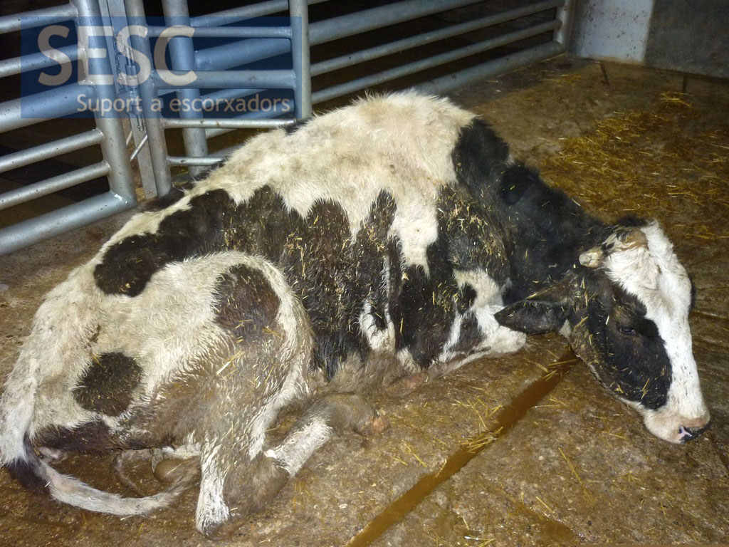
The cow was dehydrated and cachectic at its entry into the slaughterhouse showed depression and had difficulty walking.
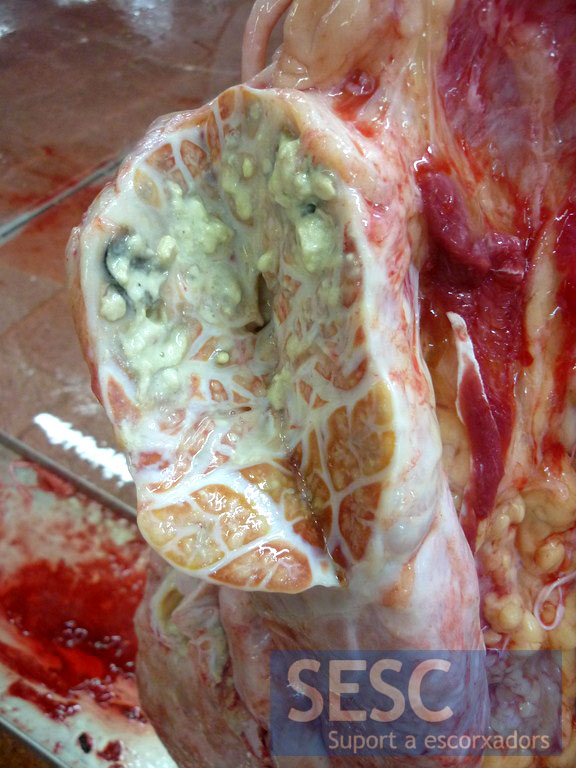
The most significant lesion was found in the lungs and consisted in fibrinopurulent abscessing pneumonia.
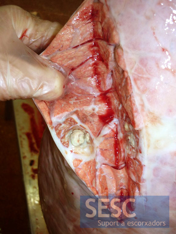
The distribution of lesions was multifocal.
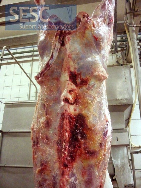
Cachectic carcass, with subcutaneous hemorrhages.
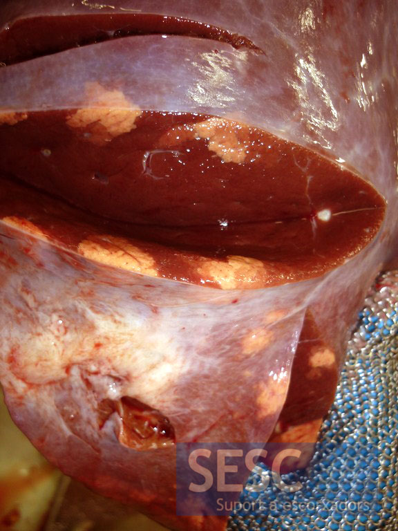
Multifocal hepatic lipidosis, this image could be confused with necrosis of the liver parenchyma.
