Infectious bovine keratoconjunctivitis in calves
In a group of friesian calves, ocular lesions were observed in 25 of them. The lesions consisted on uni or bilaretal white discoloration of the cornea that, in some cases, also presented ulcerated areas.
This lesion consists of keratoconjunctivitis (inflammation of the cornea and conjunctiva) with corneal ulcers. The histopathological study was also able to identify hyphema (presence of hemorrhage the anterior chamber of the eye).
Meat inspectors suspected of an infectious etiology, among the differential one must include Moraxella bovis, but also other agents such as herpesvirus (Malignant Catarrhal Fever or infectious bovine rhinotracheitis, although other lesions should be observed), Mycoplasma and fungi (maybe not in so many animals).
Swabs were taken from lesioned corneas in 6 animals for microbiological culture and in 2 of them colonies compatible with Moraxella were isolated. Additionally, PCR was used to rule out Mycoplasma bovis (the six swabs were negative).
This disease is known as infectious bovine keratoconjunctivitis (pinkeye) caused by Moraxella bovis. It is more common in the summer due to the greater presence of flies that act as vectors for the bacteria between animals and is usually aggravated by secondary infections and poor environmental factors (dust, sunlight ...).
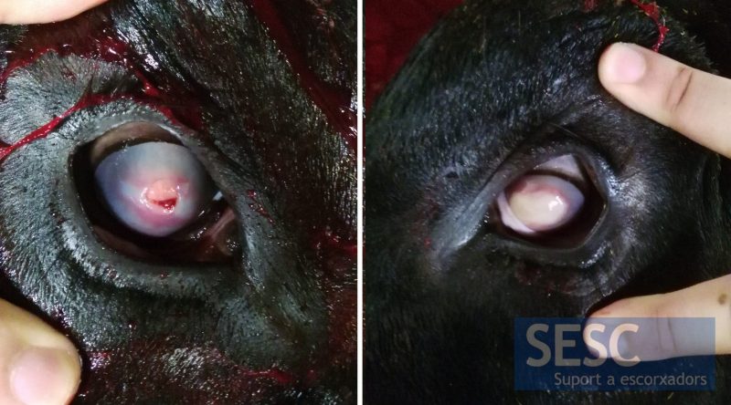
Appearance of the ocular lesions observed in calves.
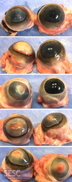
Different degrees of severity of lesions submitted. In some animals it was bilateral and, in others, unilateral. In some cases the cornea was completely ulcerated.
