It is not always a lymphoma
In a 2 year old, female, pig carcass, multiple whitish, hardened nodules were observed in the liver and mesenteric lymph nodes. The cut surface was pink with whitish margins.
The inspectors immediately suspected of a neoplasia of lymphoid origin and submitted formalin fixed samples to confirm the diagnosis.
We must confess that all of SESC pathologists agreed with the diagnosis of the inspectors and what we saw later, under the microscope, cought us by surprise...
Histologically, the nodules of the liver and mesenteric lymph nodes, were formed by abundant fibrous tissue, an indicator of the chronicity of the lesion, with a granulomatous inflammatory infiltrate with abundant eosinophil polymorphonuclear leukocytes and multinucleated giant cells caused by the presence of fungal hyphae. It consisted therefore of a chronic fungal hepatitis and lymphadenitis.
It is a rare lesion, probably caused by fungi of the class of the Zygomicetes (including genres such as Mucor, Rhizopus or Cunninghamella, among others). These fungi are found in the environment and can enter through skind wounds, is not a contagious disease. In this case, involvement of the mesenteric lymph nodes may suggest a digestive entry. The disease caused is known as Zygomycosis or mucormycosis. In the absence of fresh tissue samples, we could not attempt to cultivate and characterize the fungus causing the lesions.
In this lesion's differential diagnosis, besides neoplasia, other agents causing granulomatous inflammation should be included such as mycobacteria and parasites, even though, macroscopically often have a different appearance.
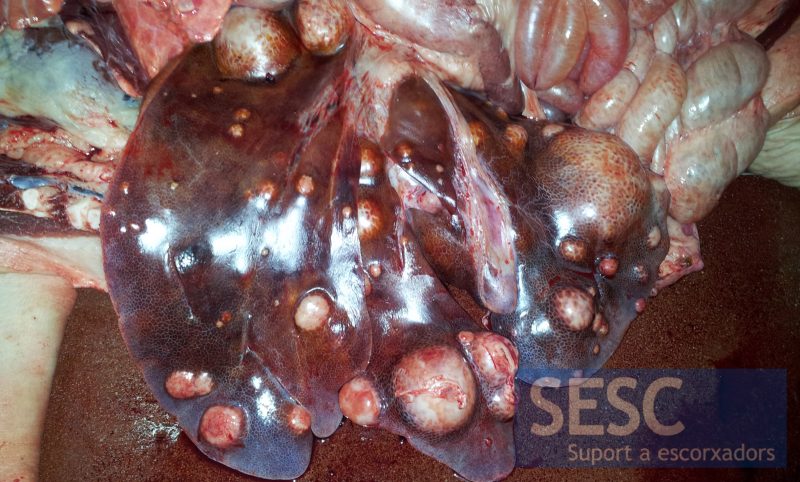
Multiple whitish nodules in the liver parenchyma.
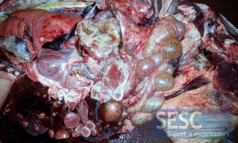
Some abdominal lymph nodes were also increased in size.
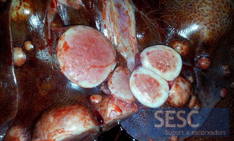
Details of the lesions once cut, the inner surface was pink, with white margins.
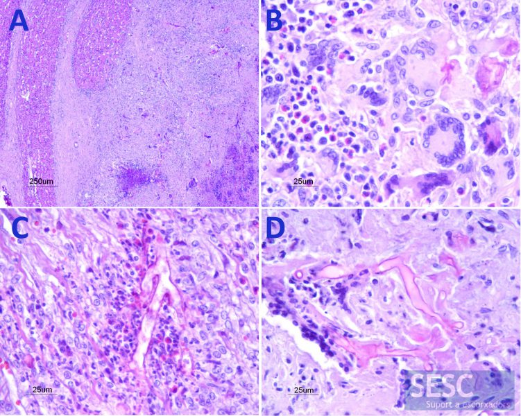
Hematoxylin and eosin staining. A: Compressed liver tissue (left) with a fragmetnof a nodule composed of abundant fibrous tissue and mixed inflammatory infiltrate. B: Details of the inflammatory infiltrate rich in macrophages, multinucleated giant cells and eosinophilic polymorphonuclear leukocytes. C and D: Presence of thin-walled, branched fungal hyphae surrounded by inflammatory cells.
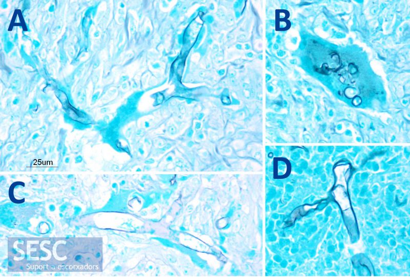
Grocott stain. A, C and D: The Grocott stains in black the thin walls of the fungal hyphae. B: Fragments of fungal hyphae phagotyzed by a multinucleate giant cell.

3 comment(s)
Comment on Prok Industry Linkedin group by Casey Bradley:
Is this not why we love science…it always tends to surprise and amaze.
Interresting article.Thanks for the information.
Thanks for reading us. We all placed out bets on a lymphoma. It was a surprise to find out it was not!