Liposarcoma in a sow
In a three years old sow carcass multinodular, whitish lesions were observed adjacent to the kidney and also affecting the renal parenchyma.
The differential diagnostic was of an adrenal gland tumor or a lymphoma.
Histopathological study showed that all masses were formed by an anaplastic mesenchymatous malignant neoplastic cell proliferation.
The tumour cells had multiple small empty vacuoles in their cytoplasm which pointed to a diagnosis of liposarcoma (malignancy arising from adipose tissue cells).
Sudan Black staining, done on formalin-fixed but not paraffin-embedded tissue, stained the content of these vacuoles confirming that they contained lipid material.
This case has been published in the scientific journal: Journal of Veterinary Diagnostic Investigation.
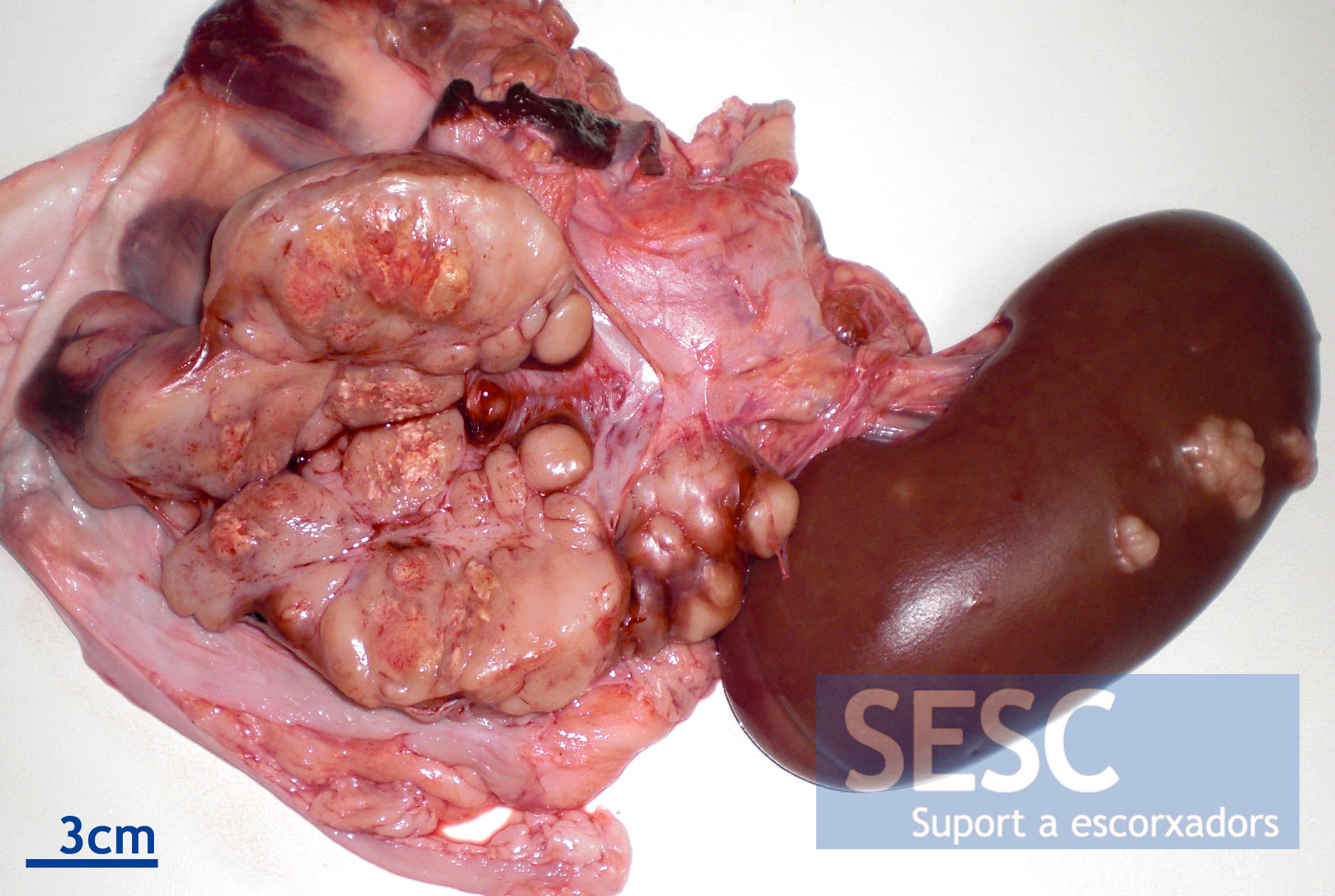
Whitish multinodular lesions adjacent to the kidney. Yellowish areas correspond to neoplastic tissue necrosis.
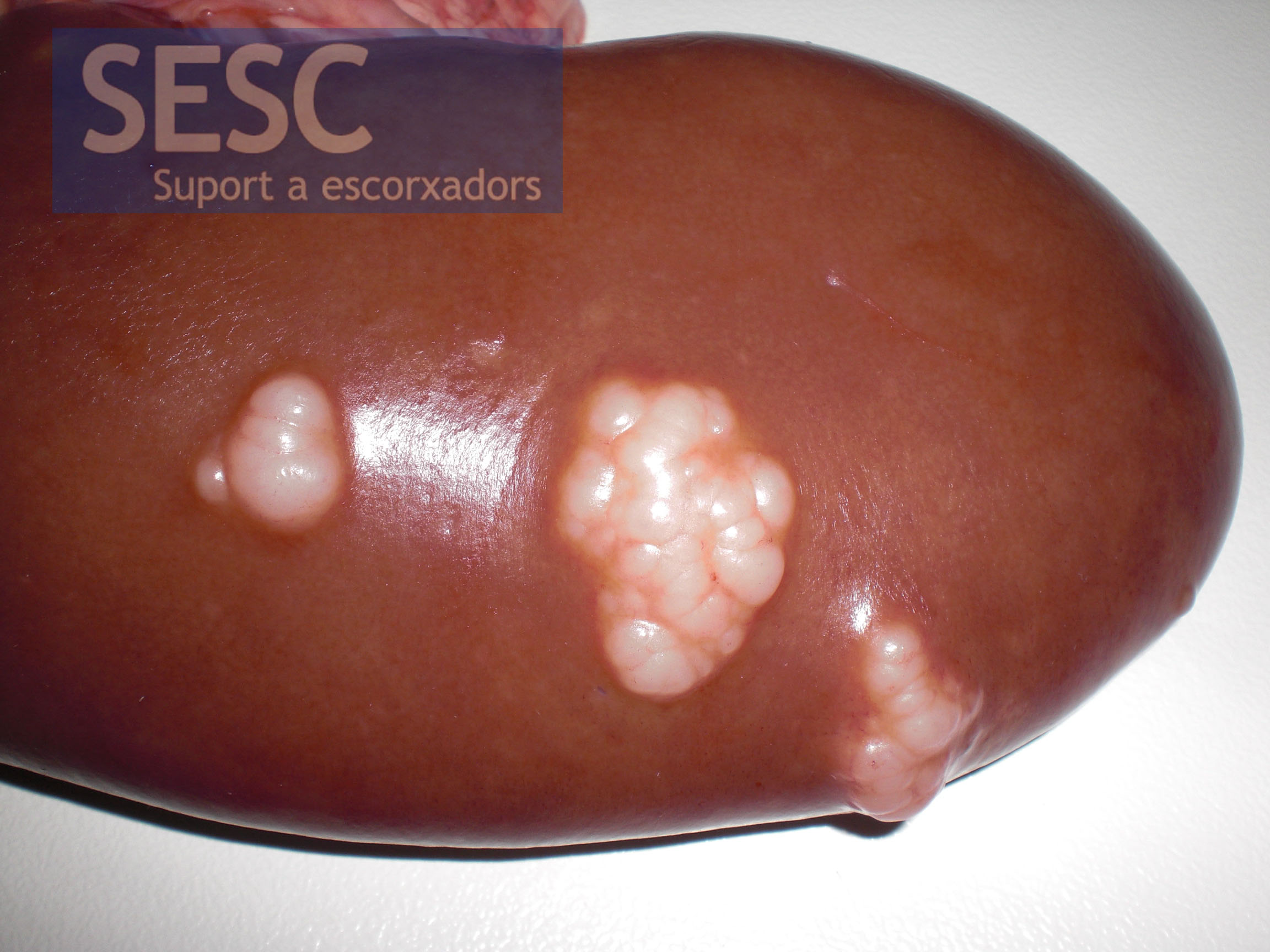
The kidney also had multiple whitish nodules.
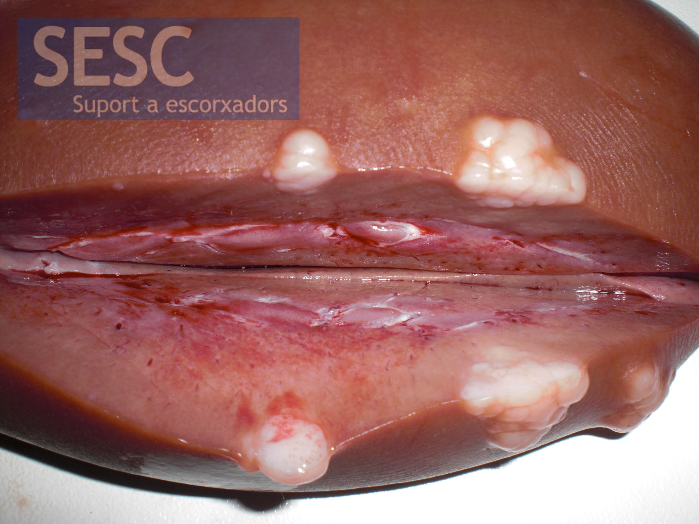
When cut the nodules extended into the renal parenchyma.
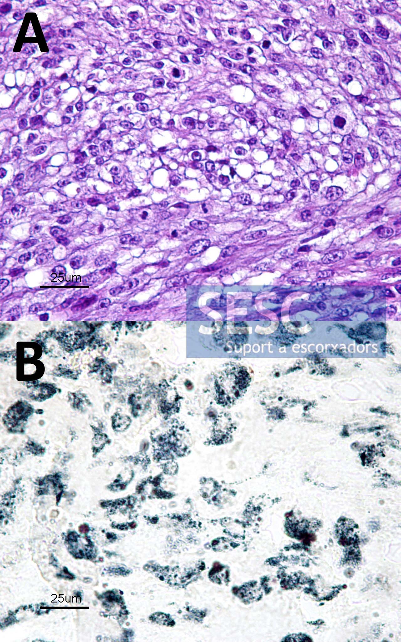
(A) Hematoxylin-eosin staining where mesenchymal, anaplastic cells can be observed with numerous mitotic images and empty vacuoles in the cytoplasm. (B) Sudan Black staining demonstrates that tumor cells bear lipid-laden droplets inside.
