08/04/2013
|
Bovine
0
Myocardial abscess in a calf
The differential diagnosis of such a lesion should include:
- Neoplasia
- Inflammatory lesions such as:
- An abscess.
- Granulomatous type lesions caused by parasites (cysticercosis, for instance) or even TB (although the location makes it unlikely to be an option).
- A more remote option would be a traumatic reticulo-pericarditis, but it would have been associated with inflammation of the pericardium.
Looking at the images it can ne seen that when cut the lesion has purulent content, so the a bacterial etiology should be considered in the first place. Pyogenic bacteria arriving to the heart though a hematogenous route. This is an unusual location for an abscess.
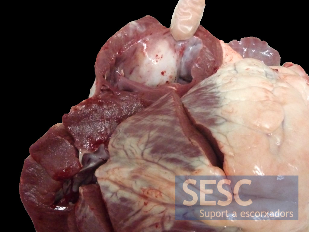
The finger points to the whitish nodule that protrudes into the ventricular cavity.
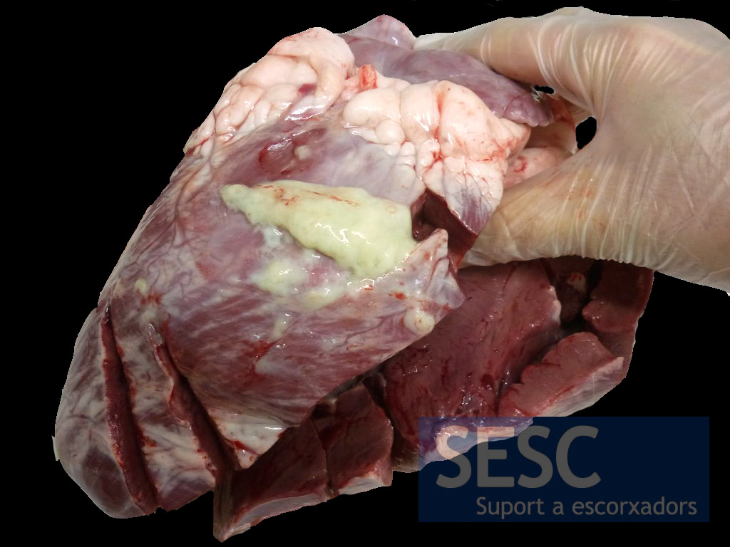
When cut, the lesion is filled up with purulent material.
