03/03/2008
|
Bovine
0
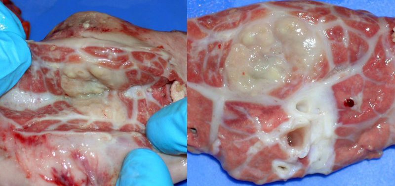
Nodular lesions in the lung parenchyma.
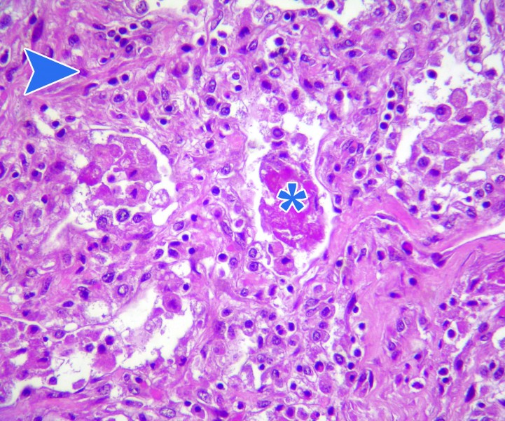
Histopathological study showed interlobular septa thickening and an area of fibrosis with multiple foci of coagulative necrosis associated with bacterial foci and presence of purulent inflammatory infiltrate. Lesions were consistent with a chronic fibrinous pneumonia caused by a bacterial agent, possibly a past resolved pleuropneumonia.
Lesions associated with bovine TB were ruled out.
One of the most common causes of bacterial pneumonia is Pasteurellosis (Mannheimia haemolytica infection (formerly known as Pasterurella haemolytica), Pasteurella multocida and Histophilus somni (formerly Haemophilus somnus)). Bacterial pulmonary complications might be secondary to a viral infection, stress, etc..


