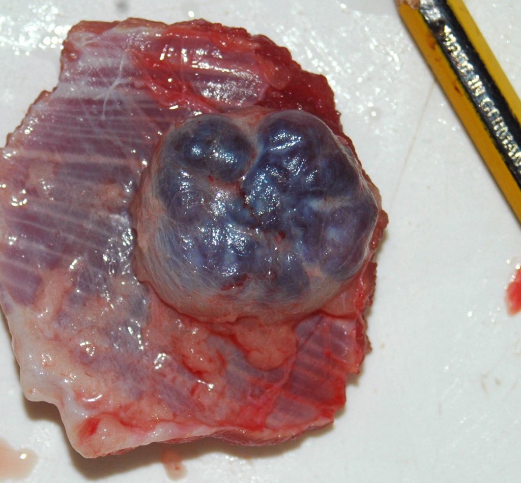
Nodule in the fascia of a 12 months old bovine.
Histopathological examination showed lesions consistent with lymphoid hyperplasia associated with intensive blood resorption, this lesion is known as hemal node.
The hemal nodes are structures that can be observed in ruminants, and rats, are formed by a fibromuscular capsule and its structure is similar to that of lymph nodes. The hemal nodes have blood circulation as well as afferent and efferent lymph vessels. They also have germinal centers but the paracortical area is reduced compared to the lymph nodes. In the subcapsular sinus (in lymph nodes only a few cells can be observed) hemal nodes usually contain erythrocytes, an amount equal to that found in the blood, these erythrocytes are hardly observed in the lymph vessels which enter and leave these nodules. The hemal nodes sinuses can be full of blood resembling the appearance of the spleen. As the spleen, macrophages occupy the trabecular areas and phagocytose erythrocytes. There hasn't been found evidence of hematopoiesis phenomena in hemal nodes.
Hemal nodes are not the same pathogenically entity than bleeding resorption in lymph nodes or lymph nodes hemorrhage, although in both cases erythrocytes and siderophages can be observed in the medullary subcapsular sinus. For example, it is relatively common to see blood resorption in the mediastinal lymph nodes due to extravasated blood during the slaughter process.


