Pyogranulomatous eosinophilic myositis by Sarcocystis spp
They measured between 0.2 and 1 cm in diameter and were observed extensively in the carcass (mesentery, masseter, heart, tongue, abdominal ...).
The histopathological study revealed eosinophilic pyogranulomas (Figure 3) associated, in some cases, to the presence of Sarcocystis spp. cysts either intact or in various stages of degeneration. Sarcocystis spp. cysts were also observed within muscle fibers, which is where they are usually seen but without creating any reaction.

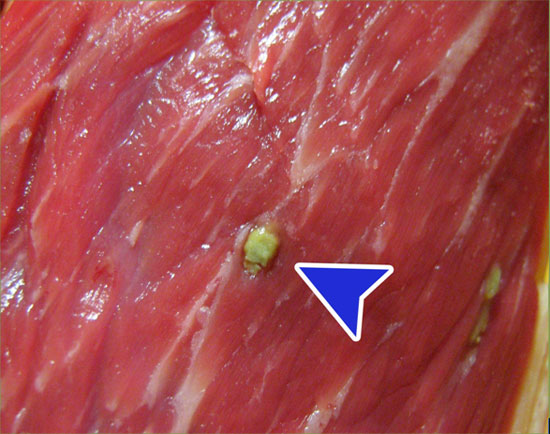
Greenish granuloma of about 1cm in diameter (arrow).

The lesion (arrow) is repeated in a generalized manner affecting limb muscles (picture) but also masseter muscles, diaphragm, heart, tongue, etc..

Microfotografía de tinción de hematoxilina-eosina. Se observa un piogranuloma en el interior del cual podemos ver un quiste de Sarcocystis spp. rodeado de una intensa reacción imflamatòria de polimorfonucleares eosinófilos. También se pueden apreciar fibras musculares en degeneración (asterisco).

