Stone-like lung lesions in a horse carcass
In a 21 years old, cross breed, mare carcass several masses with a very hardened consistency (stone-like) and considerable size (10-15 cm) were observed located in the right apical lobe and in the ventral margins of diaphragmatic lobes.
Histopathological study showed multifocal lesions of granulomatous, chronic, inflammatory nature. These lesions developed areas of extensive fibrosis with mineralization or osseous metaplasia (i.e. bone tissue that is generated in the center of the fibrous lesion, which explains the hardness of the lesions found on the carcass).
A series of tests were performed to determine the cause of these inflammatory lesions:
- The Ziehl Neelsen staining was unable to determine the presence of mycobacteria. Both PCR and direct mycobacteria culture also gave negative results.
- The Gram stain did not allow to observe gram positive bacteria.
- Neuther did Groccott's stain allow to observe fungal structures.
Thus it was not possible to determine the etiology of these lesions probably because of their advanced chronicity preventing to identify the cause. A neoplastic process was ruled out.
In the differential diagnosis an equine multinodular pulmonary fibrosis was included, it is a disease associated with equine herpesvirus 5 infection. Again, the chronicity of the lesion makes it difficult to associate it to this entity.
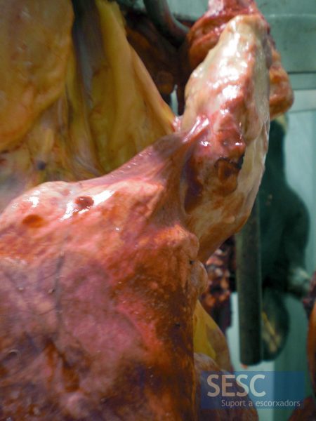
Lung lesions.
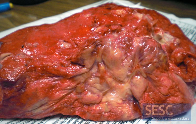
Lung lesions.
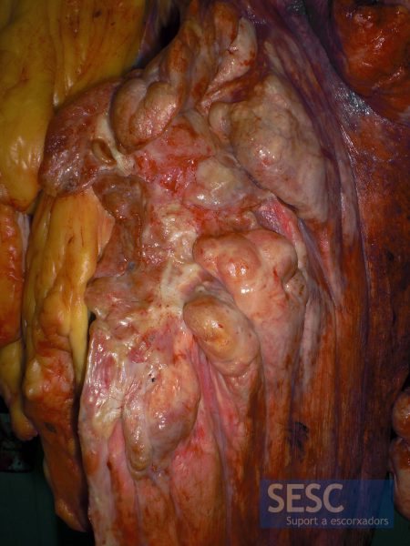
Lung lesions.
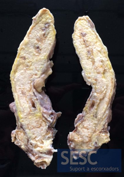
Longitudinal sections of one of the masses after decalcification.
