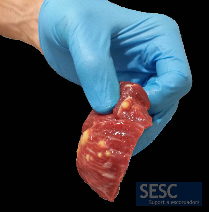
Whitish nodular lesions in the muscles of a cow
Even though, macroscopically, these lesions were indeed compatible with typical Cysticercus bovis parasitic granulomas, microscopic observation showed that it consisted of severe multifocal embolic-metastatic myositis. That is, purulent-necrotizing lesions with presence of bacteria most likely originated in a (non reported) primary focus in another location, such as a bacterial valvular endocarditis or a suppurative lesion in some other point of the carcass.
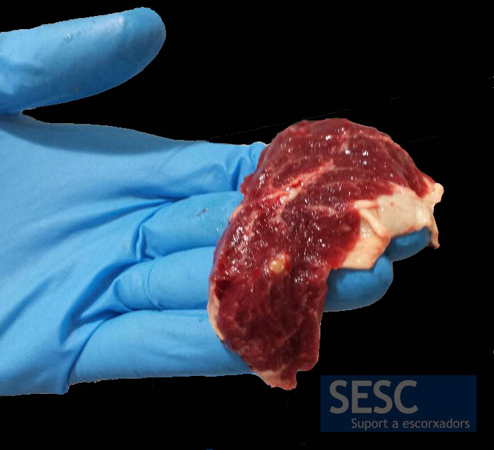
Yellowish nodular lesion in the masseter muscle.
In the myocardium multiple yellowish-white lesions were observed.
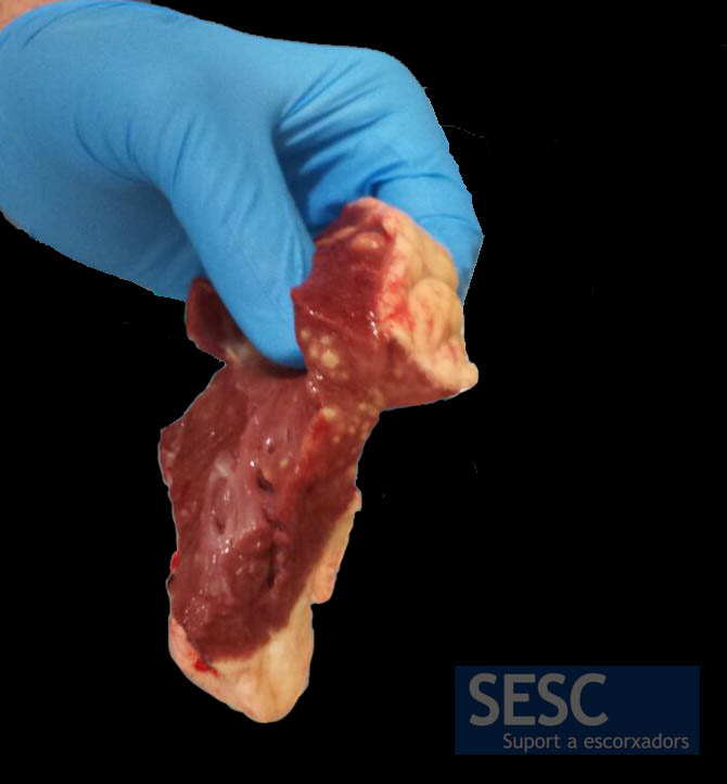
The diaphragm also showed multiple nodular lesions.
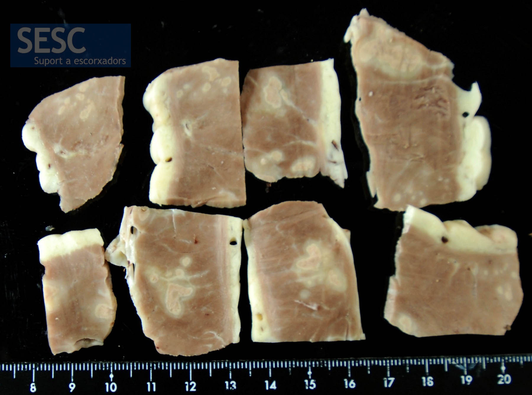
Sections of muscle tissue, fixed in formaldehyde, where the lesions can be observed.
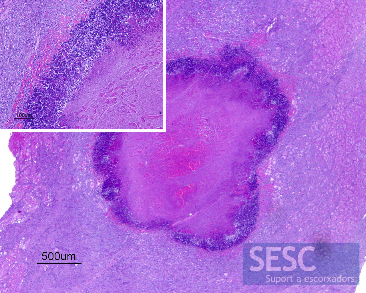
Photomicrograph of hematoxylin-Eosin staining. Muscle lesion with central necrosis surrounded by inflammatory suppurative infiltrate and macrophagic-lymphocytic reaction with evident vascular activation.
