Hepatic cirrhosis in a calf
Histopathological examination evidenced bile duct hyperplasia, and an evident increase of conjunctive tissue in portal spaces and in a mesh of fibrovascular septa that completely surrounded the hepatic lobuli. The nodules corresponded to islands of residual liver tissue regeneration. A morphological diagnosis of hepatic cirrhosis was established.
Cirrhosis is the final, irreversible, phase of various pathogenic processes leading to hepatocyte cell death (apoptosis or necrosis) and fibrosis with chronic active inflammation. Among these processes one can find intoxication and cirrhosis due to parasitic infestation.
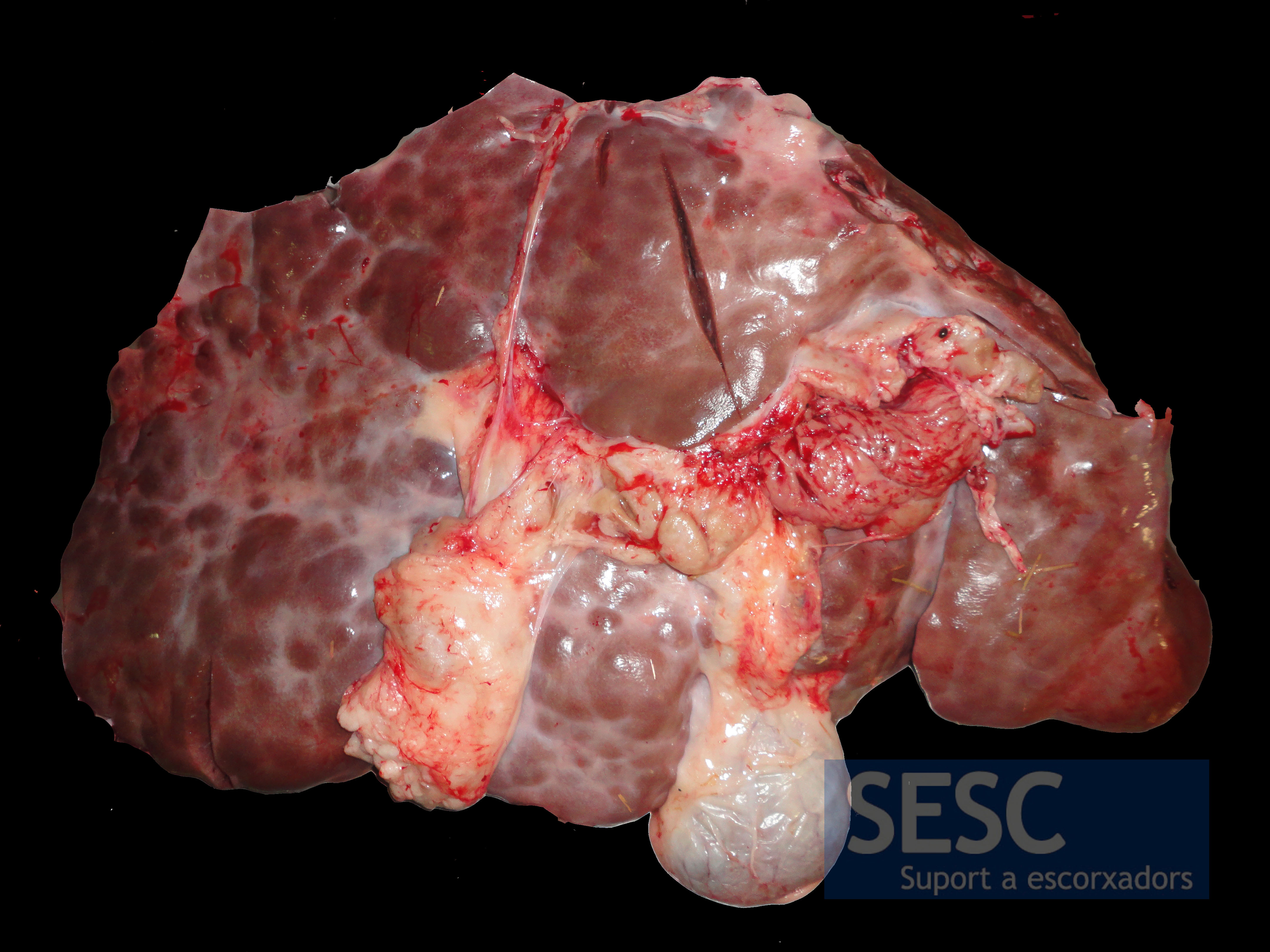
Multiple nodular lesions separated by paler tissue.
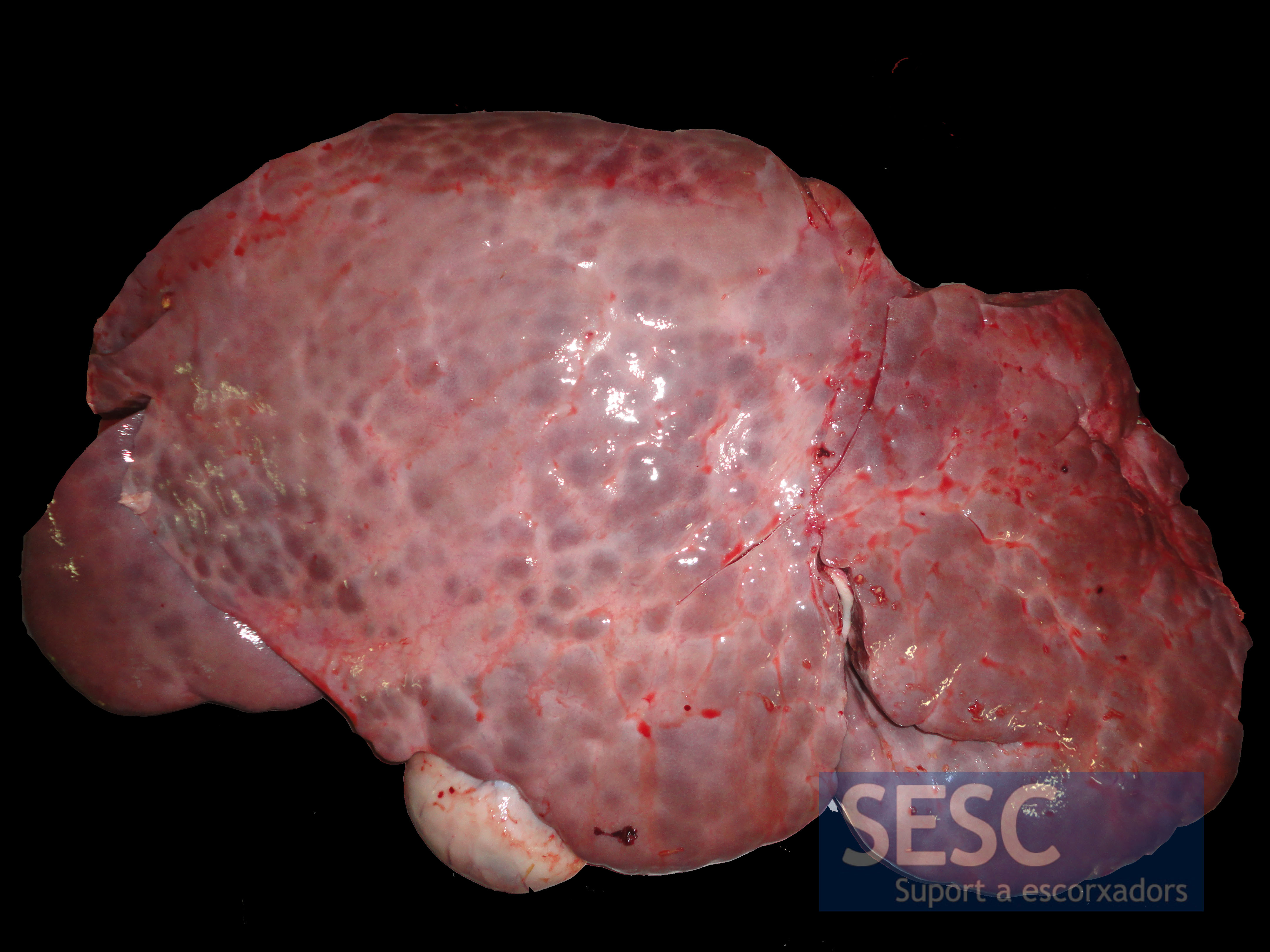
View of the diaphragmatic surface of the liver.
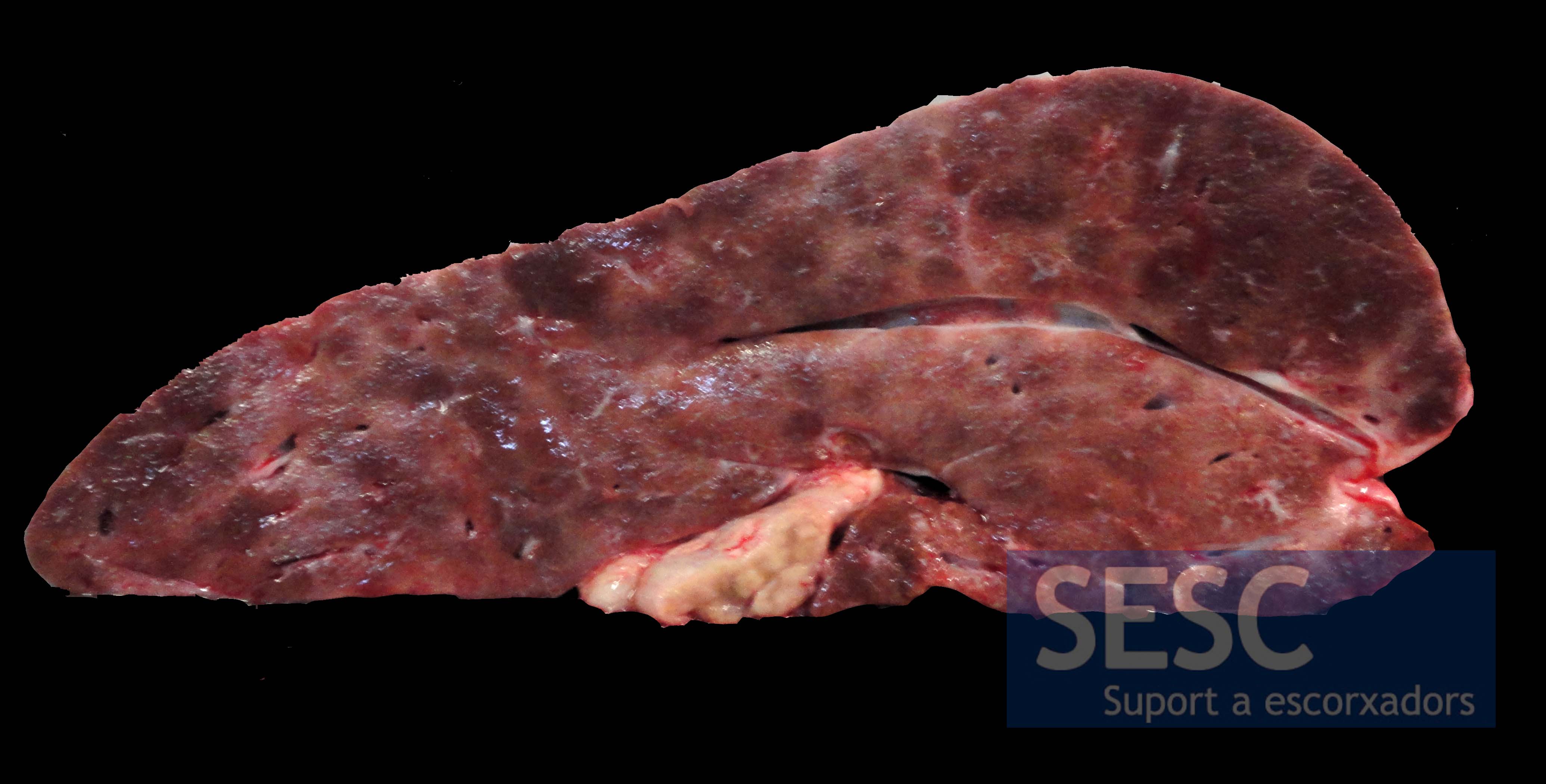
The lesion can also be observed when the parenchyma is sectioned.

3 comment(s)
Hola, tengo algunos terneros que tienen fotosensibilidad. Al parecer eczema bovino. Que sugieren para curarlos?
Tengo un toro que se lleno de agua la pansa y le puse unas agujas y le salió mucha agua . El veterinario me dijo que no tiene cura .
Quisiera saber que es eso
El acúmulo de liquido en la cavidad abdominal se conoce como ascitis y puede tener múltiples causas. Desde procesos inflamatorios (peritonitis), hemorrágicos, neoplasias, etc. Es necesario determinar que tipo de liquido se acumula. Una patología hepática, como la cirrosis que se observa este caso, puede cursar con hipoalbuminhemia lo que provoca una disminución de la presión oncótica en al sangre y por eso sale fluido al exterior de los vasos. También sucede en casos de malnutrición extrema.