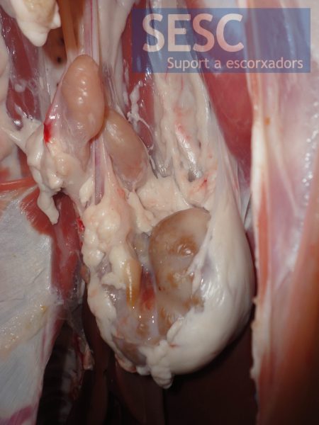
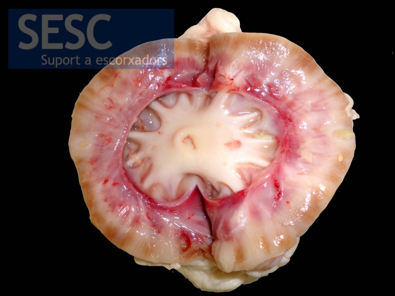
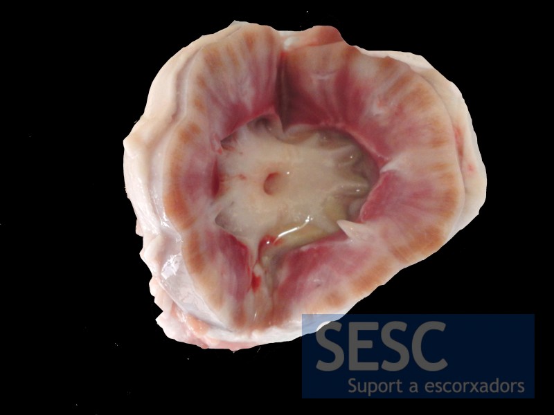
Chronic pyelonephritis in a lamb
When sectiones the pallor of the renal parenchyma was also appreciated as well as an apparent loss of renal cortex tissue.
Histologic lesions observed were of a mixed chronic inflammatory infiltrate consisting of macrophages, neutrophils, lymphocytes and plasma cells, distributed through the renal cortex and medulla, both in the interstitium and in the lumen of the tubules. Glomeruli showed little or no lesion at all. There was a widespread fibrosis at its initial stages.
This lesion is called chronic pyelonephritis and probably has a bacterial ascending origin, i.e. originated in the lower urinary tract. The smell of ammonia from the carcass is indicative of a state of uremia secondary to a renal failure associated with the observed lesions.


