30/10/2012
|
Porcí
2
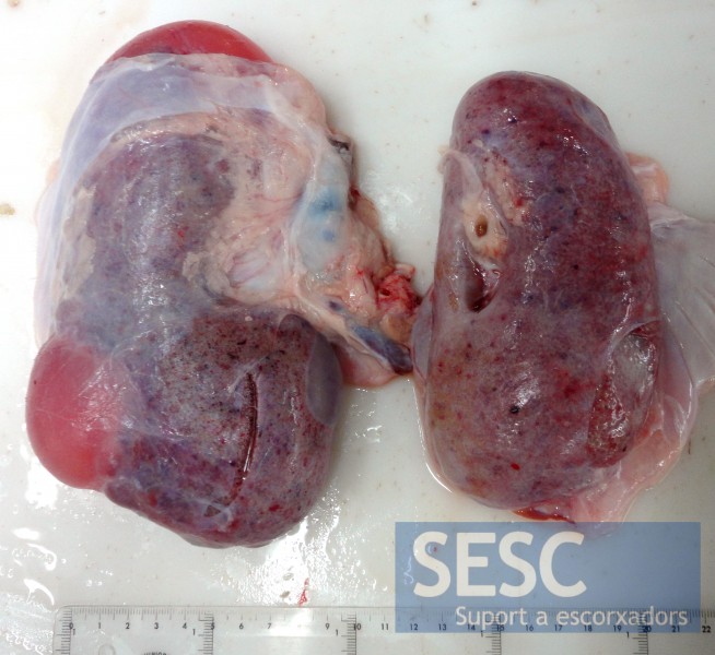
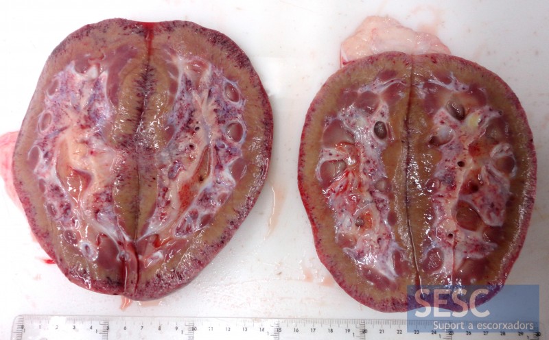
Petèquies renals en un porc
L’estudi histopatològic evidencia la formació de múltiples lesions cavitàries al parènquima de l’escorça renal. També evidencia altres lesions compatibles amb una síndrome de dermatitis i nefropatía porcina(SDNP) en fase de resolució (glomerulonefritis necrotitzant proliferativa). La SDNP sol afectar també a la pell (veure entrades Lesions cutànies hemorràgiques i SDNP), però no sempre és el cas.
Mitjançant hibridació in situ es va descartar la presència de genoma de circovirus porcí tipus 2, que s’ha associat a aquesta malaltia.


2 comment(s)
En aquest cas i en un altre de semblant l’inspector que ha remès les mostres comenta que observa un canvi de consistència en el greix perirrenal. Aquest apareix més fluid i regalima per la canal.
Histopatològicament només s’aprecia un lleuger component inflamatori.
Algú ha observat aquest tipus d’alteració en casos de SDNP?
En este caso y en otro parecido el inspector que remite las muestras describe un alteración de la consistencia de la grasa perirenal: esta aparece más fluida y rezuma por la canal.
Histopatológicamente solo de aprecia un ligero componenete inflamatorio.
¿Alguien ha observado este tipo de alteración en casos de SDNP?
In this case, and also in a similar one, the meat inspector described an altered consistency of the peri-renal adipous tissue. The fat appeared somwhat liquefied. Under the microscope only a mild inflammatory componenet was observed.
Has anyone seen such changes in PDNS cases?
The following comment was recieved through LinkedIn groups:
What is the incidence of this pathological finding? I have seen just a few cases in pigs death at farm or at slaughterhouse. In chronic forms of Exudative Epidermitis you also can find this damage. Both are not usual to find. Also I have seen severe renal damage in cases of some antibiotics over dosification.
Out pathologist answer was:
Thanks for your question. This is a rare lesion, and we have not seen this kind of lesion before. It belongs to a pig slaughtered at the normal age, arriving without signs to slaughter. We have classified it tentatively as a chronic case of PDNS based only on the presence on some glomeruli of fibrin plugs in the Bowman’s space. We concluded it is a PDNS case in resolution phase. Some glomeruli and glomerular vessels had thickened basal membranes (PAS staining), but there was no clear inflammatory changes in the kidney at this stage, which is somehow unexpected for a chronic PDNS. We performed in situ hybridization for PCV-2 in the kidney and in the submitted lymph node, with negative results.
The most striking difference of this case with other PDNS chronic cases that we have seen is the externes lesion in the outer cortical layer of the kidney. There were multifocal areas of absence of the normal renal tubular structures, replaced by empty irregular cavities (NOT cystic), covered by flat epithelial cells, and with the only presence of glomeruli, lymphoid follicles, and vessels with hyperthrophic walls (very few islands of tubular structures). Some of these cavities were filled with plasma-like fluid, but other had blood. The lesion was clearly NON cystic in nature, and these cavities appear to be the result of loss of renal cortical tissue (isquemic?, toxic?).
We can share histo slides of this case if you have interest on the histopathology of the lesion.