Pig carcasses with black guts
Samples were submitted of pig intestines from a batch in which 100 animals showed a black coloration of the intestinal mucosa along its entire length. The coloration was limited to the mucosa without delving into the other layers of the intestine. No lesions were reported in other viscera.
The histopathological study showed the presence of a moderate number of macrophages at the level of the mucosal lamina propria that contained a granular pigment of brown coloration. Occasionally free pigment could also be observed on the lamina propria itself. In the lymph nodes some macrophages with the same pigment could also be observed.
PEARLS staining was performed to detect iron which evidenced the presence of hemosiderin in some macrophages, but it was not the main component of the accumulated pigment. A copper stain with rubeanic acid was also performed, with a negative result.
It is therefore an accumulation of an inert pigment (does not generate an inflammatory response) of very probable dietary origin, but its composition and origin are unknown. Lipofuscinosis is ruled out because in this case the accumulation would be in the smooth muscle fibers and not in the lamina propria.
If you have seen similar cases or know of pigments that, introduced in the diet, can give this alteration, we invite you to share it in the comment section at the end of the entry.
In humans a condition named Melanosis coli has been described, a deposit of pigment in macrophages of the lamina propria also described in pigs.
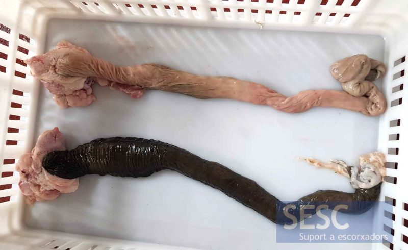
Black coloration of the intestinal mucosa (bottom) compared to a normal intestine (top) (the intestine has been turned upside down to allow observation of the mucosa).
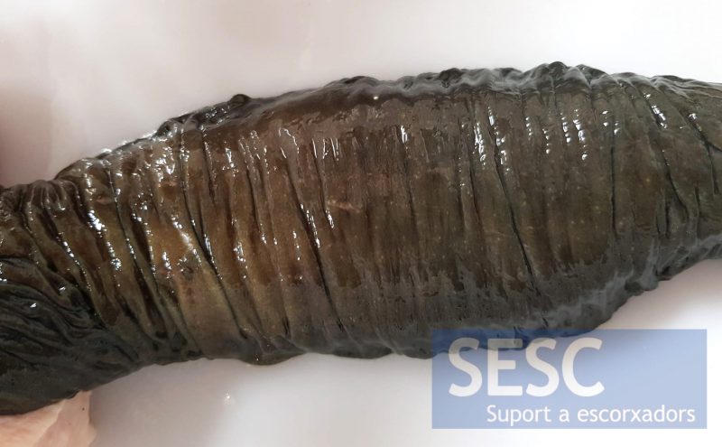
Detail of the coloration of the mucosa of the large intestine.
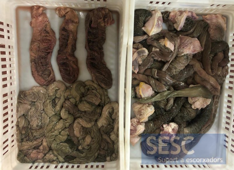
The intensity of the change in coloration varies between animals and in the different sections. In some cases the hue is greenish.

1 comment(s)
It might be a case of Melanosis coli:
https://journals.sagepub.com/doi/pdf/10.1177/0300985814559403