Sheep can also have tuberculosis
In this blog we have previously seen cases of carcases with tuberculous lesions in the bovine, caprine and wild boar species. In this post we want to show images of tuberculous lesions in the ovine species.
Ovine, in spite of being susceptible to infection by mycobacteria that cause tuberculosis (Mycobacterium tuberculosis complex) all of them zoonotic (can cause tuberculosis in humans), are not included in the plan for the eradication of bovine tuberculosis and no routine tests are performed to control them. Therefore, inspection in the slaughterhouse becomes a key tool for detecting outbreaks of this disease in the ovine species.
Recently, this case has been published in which tuberculosis transmission was shown between goats and sheep from the same farm, reinforcing the idea that these species of small ruminants should be considered as domestic tuberculosis reservoirs.
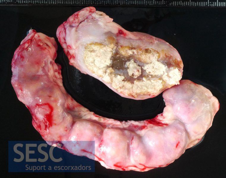
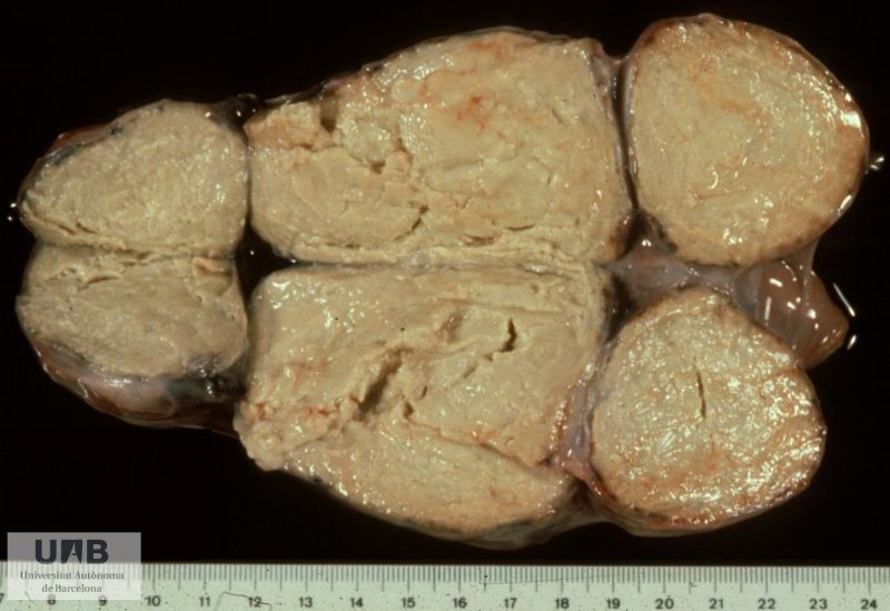
Granulomatous lesions in the mediastinal lymph nodes of a sheep. Mycobacterium caprae was isolated from the lesions.
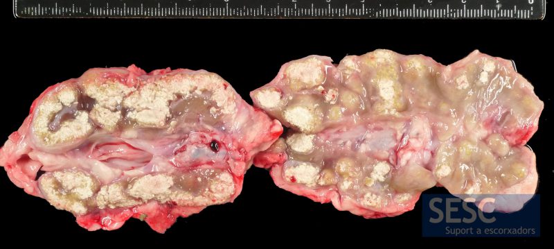
The lesions were hardened and had a gritty consistency when cut (mineralization).
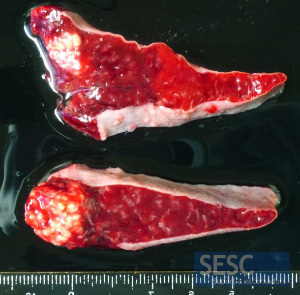
The spleen also had granulomatous lesions.
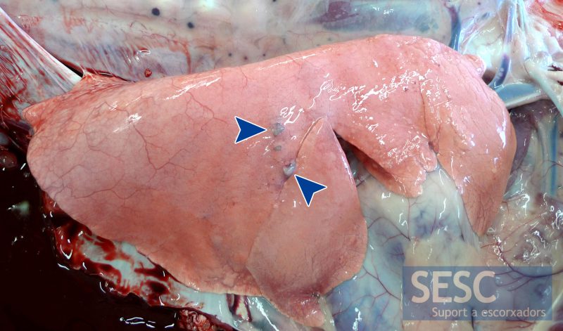
Sub-pleural tuberculosis granulomatous lesions in the lungs of a sheep (experimental infection with Mycobacterum caprae performed at IRTA-CReSA).
The main differential diagnosis when we find caseous lesions in the lymph nodes of a sheep carcass is pseudotuberculosis, also known as "caseous lymphadenitis" caused by Corynebacterium pseudotuberculosis. These lesions are usually well encapsulated and the caseum has a characteristic concentrical appearance that is not usually observed in tuberculosis, in these cases, it is also important to rule out tuberculosis. You can see more images in the SDPV image database.
