15/09/2015
|
Bovine
6
Black subcutaneous lesion in the neck of a calf
Histologically an accumulation of amorphous, acellular and eosinophilic material was observed surrounded by a fibrous reaction and abundant macrophages loaded with intracytoplasmic granules of brown pigment. The lymph node histological appearance was normal. This lesios is compatible with a foreign body granuloma, probably resulting from the percutaneous administration of a pharmacological product, most likely oily in nature.
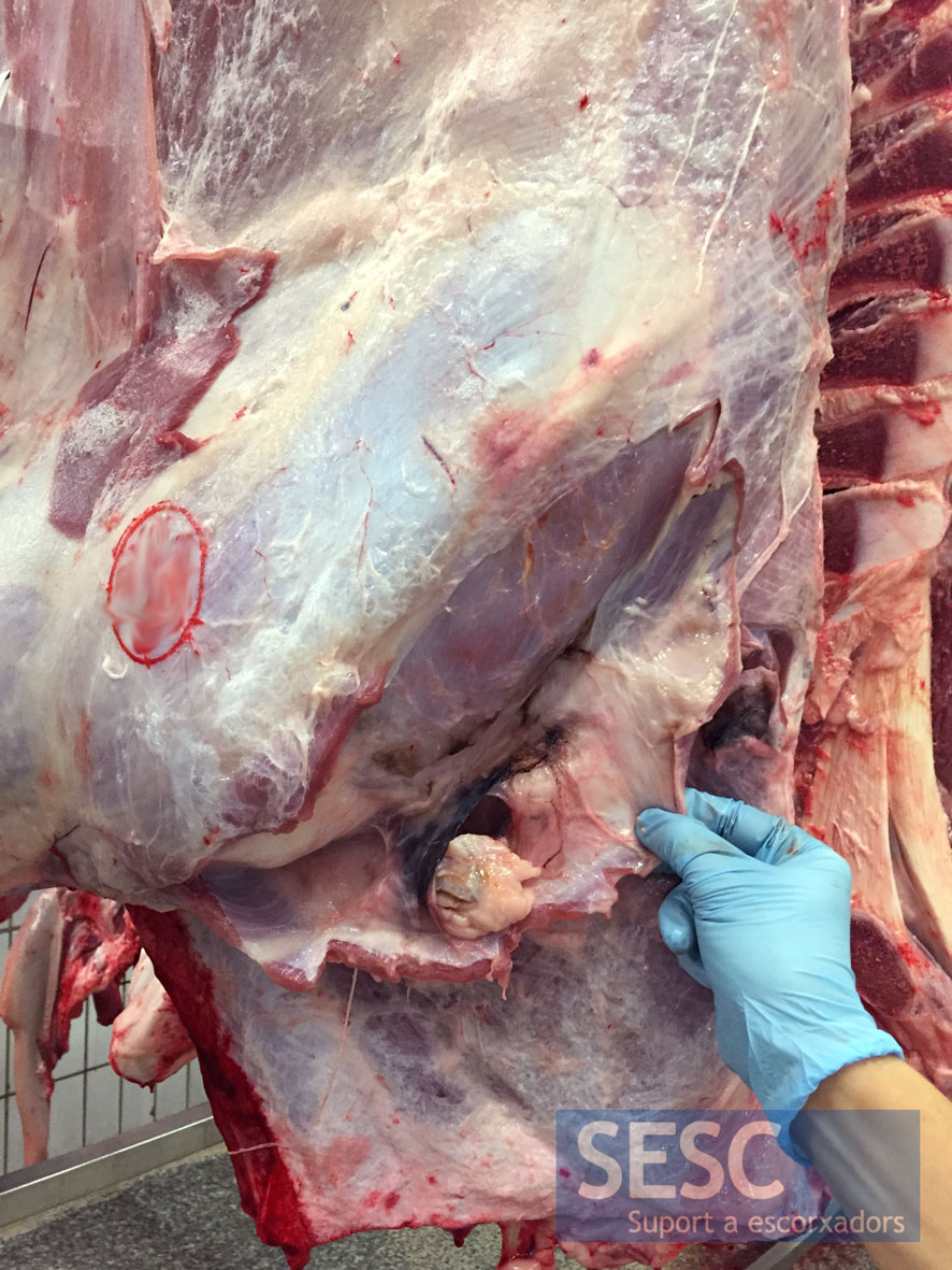
The lesion of blackish coloration, was located in the caudal cervical region close to the preescapular lymph node.
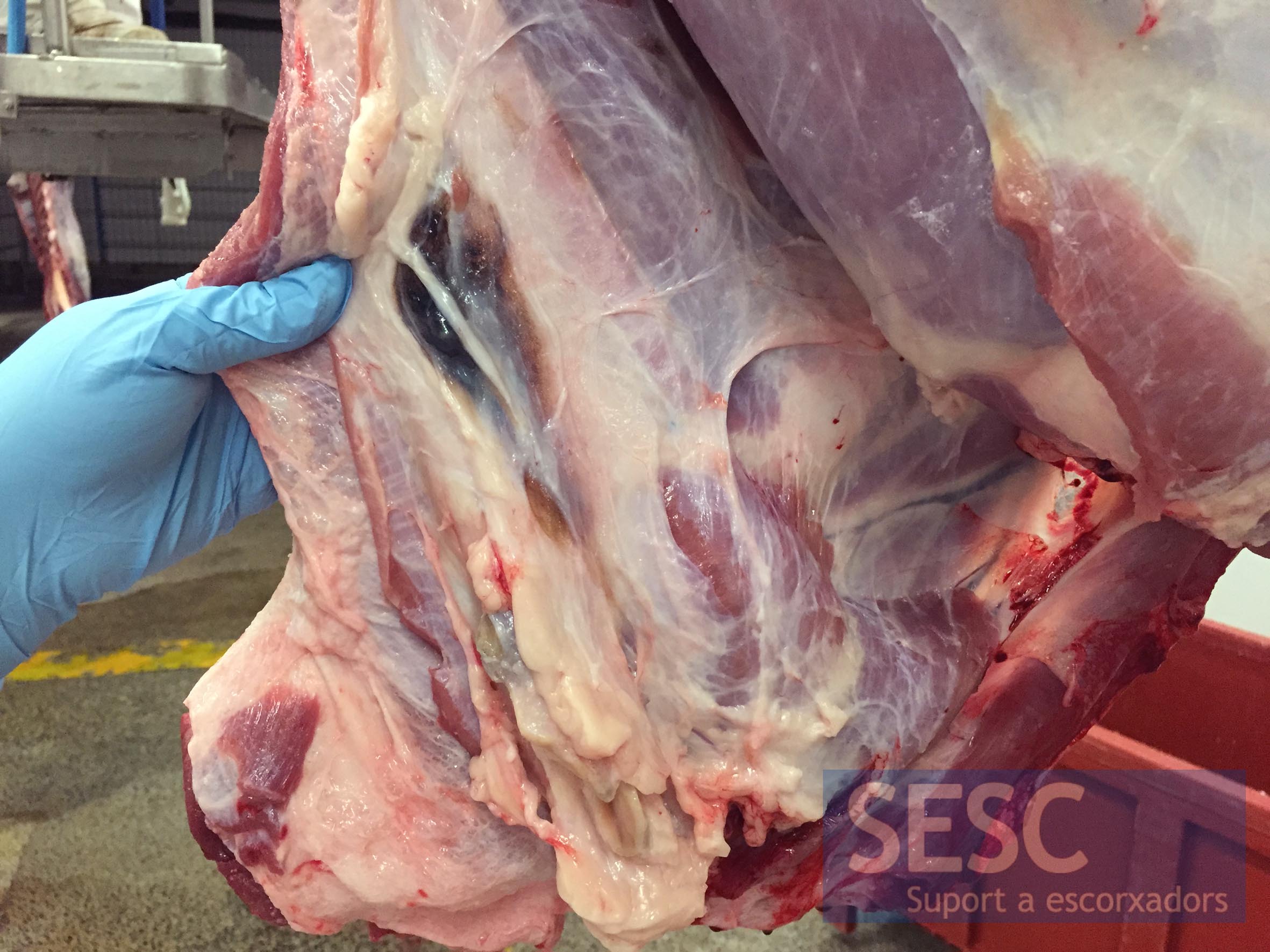
Appearance of the lesion in the connective tissue.
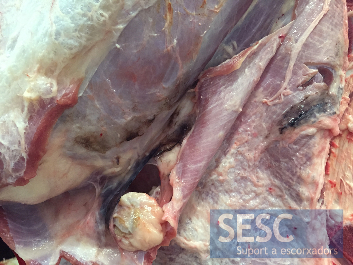
Closer look at the blackish material. Mild hyperplasia of the preescapular lymph node

6 comment(s)
Comment by Dr. Vinay Lomash at “Veterinary Pathology” Group in LinkedIN:
Clostridium novyi infection
Comment by Olakunle Tiamiyu at “Veterinary Pathology” Group in LinkedIN:
Could it be Clostridium or injected deposit of a drug?
Comment by Chretien Gielen at Veterinary Pathology Group in LinkedIN:
Iron injections are not unusual in veal calves, I am a practitioner and not very familiar with the histological findings, but an iron injection (ferrodextran) could cause the brown granules.
Best regards, Chretien
I totally agree with the histology being consistent with the diagnosis of an injection site reaction or foreign body granuloma. The brown amorphous granules are a bit unusual, although I have seen tetracyclines to this. Brown is more common with hemorrhage/hemosiderin either within an injection site or due to direct trauma. Most vaccines will have amorphous blue material- adjuvant, and other pharmaceutical agents, refractile material, basophilic or if steroid containing, acicular clefts. Any idea what possibly could have been injected? Best regards,
Dave
Thanks for your comment, Dave.
The impression of the pathologists is that the granules were of a lipidic nature (an oily excipient maybe). Tetracyclines have been suggested as a possibility or iron injection (less likely, in my opinion, since the granules have a different microscopic image),
Sadly we had no feedback on what had been injected in this case.
Cheers!
Comment by Chris Petzel at Ruminant Health, Nutrition and Production Professionals group in LinkedIn:
Depot injection of oxytetracycline?