The curious case of the yellow rabbit
We received an inquiry regarding a carcass of a 15-month-old hybrid rabbit, which the official meat inspectors condemned due to an intense yellowish coloration of the carcass. The official meat inspectors’ suspicion was hepatic jaundice, and they requested ruling out a possible case of Rabbit Hemorhagic Disease.
The carcass certainly presented a very intense yellowish coloration which was predominantly affecting the subcutaneous and perivisceral adipose tissue. Additionally, it was particularly evident because the animal had abundant reserves of adipose tissue in comparison with the rest of the animals in the batch. However, this coloration was not observed in the remaining soft tissues, in particular when the trachea and aorta were opened, they showed a normal white coloration. Different samples were collected for their study, including 1) a liver sample which was submitted to the reference laboratory (LaSAC) to rule out Rabbit Hemorrhagic Disease (which could explain hepatic jaundice), however it resulted negative. 2) fresh sample of adipose tissue for the alcohol-ether test, which resulted positive for carotenoid pigments (ruling out the presence of bilirubin, hence ruling out jaundice) and 3) samples fixed in formalin for histopathology (including liver, abdominal wall, heart, kidney, lung and lymph nodes). The histopathological study did not reveal significant alterations aside from a mild (and unspecific) presence of inflammatory cells in the liver.
The absence of significant hepatic lesions, the negative result of the ELISA against the virus of the Rabbit Hemorrhagic Disease, and the absence of bilirubin in the adipose tissue, completely ruled out that this was a case of hepatic jaundice. As the alcohol-ether test resulted, this hyperpigmentation was due to the presence of carotenoid pigments deposited in the adipose tissue.
Criteria for distinguishing jaundice from pigment accumulation: The carotenoid pigments are lipophilic and predominantly accumulate in the adipose tissue, while bilirubin has a special affinity for elastic fibres. Therefore, it is important to open the trachea and aorta (rich in elastic fibres) when suspecting jaundice: they will present an intense yellowish coloration in cases of jaundice, while showing little or none affection in cases of hyperpigmentation by carotenoids.
As in other species, the hyperpigmentation of the carcasses is related with the ingesta of carotenoids-rich feeds. However, a mutation of the gen BCO2 (encoding for an enzyme implicated in the metabolism of carotenoids) has been identified in rabbits as a cause of hyperpigmentation of the carcasses, with a mendelian recessive pattern of inheritance. In the intensive production systems the appearance of dietetic hyperpigmentation, is improbable so it is possible that this case was caused by this genetic alteration, although this was not checked. (AC)
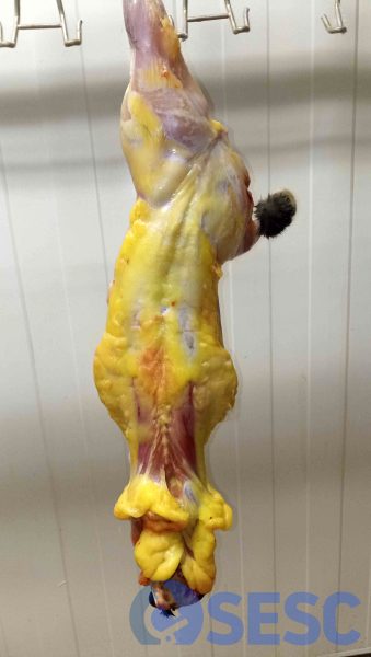
Rabbit carcass. The subcutaneous adipose tissue presents a marked yellowish coloration.
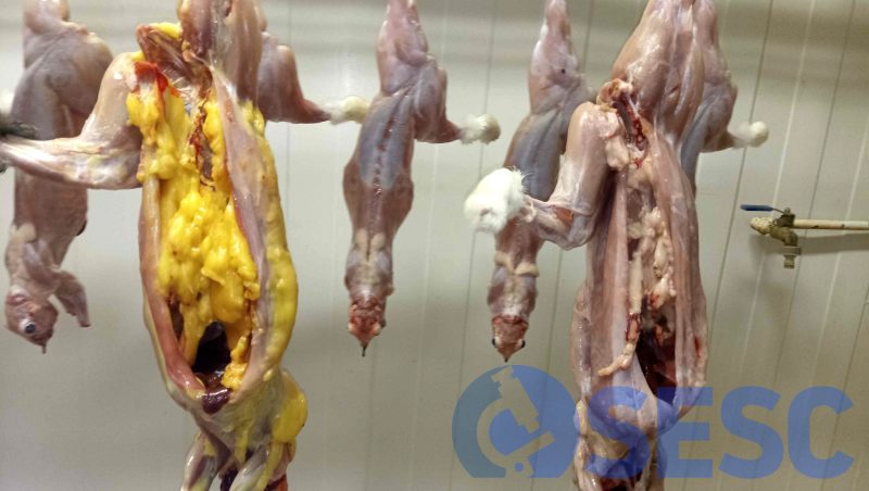
Rabbit carcass. The perivisceral adipose tissue shows a similar yellowish coloration. This change is specially evident when compared with the other carcasses, moreover this carcass presented a higher quantity of adipose tissue.
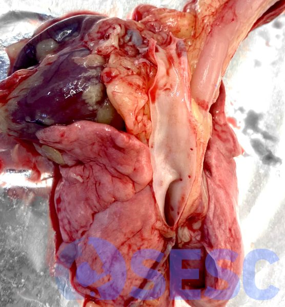
Thoracic viscera of the rabbit carcass. A yellow coloration of the mediastinal adipose tissue can be observed, however once the aorta is opened, it shows a normal coloration. This change is suggestive of hyperpigmentation due to carotenoid pigments, while in jaundice cases the aorta acquires a yellow coloration due to bilirubin having high affinity for elastic fibres. Although it is not shown in the Picture, the tracheal mucosa behaves similarly.
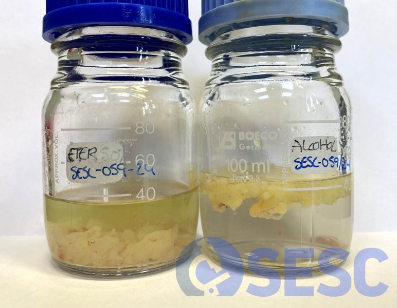
Alcohol-ether test performed with the adipose tissue of the affected carcass. After two hours, the carotenoids pigments have diffused to the ether. In contrast, the absence of coloration in the alcohol test rules out the suspected jaundice (bilirubin is alcohol-soluble).
