Rabbit hemorrhagic disease in a batch of rabbits.
We received an inquiry regarding a batch of 811 rabbits, in which the mortality in the antemortem surveillance was 2,7%, with presence of epistaxis (blood exiting the nasal cavity). At the post-mortem examination, haemorrhagic and/or icteric carcasses were observed, as well as liver alterations (reddish or pale discoloration, increased in size), and haemorrhagic lesions in the lungs and kidneys. All these findings raised the suspicion that this was a case of rabbit haemorrhagic disease.
Rabbit haemorrhagic disease is a very severe and systemic disease, caused by a calicivirus that infects lagomorphs. It usually appears as an acute haemorrhagic disease, characterized clinically by epistaxis and sudden death. Due to its rapid course, it is unlikely that animals presenting acute infection reach the slaughterhouse. If confirmed, the rabbit haemorrhagic disease is to be notified due to its severity and repercussions in the rabbit industry.
Among the submitted samples, a pale and friable liver with a lobular pattern was seen. The lungs displayed multifocal reddish areas. In one of the carcasses, the kidneys presented a marked increase in size, with an intensely darkish coloration. Samples were fixed for histopathological studies, as well as fresh liver samples for the detection of the calicivirus that causes rabbit hemorrhagic disease by PCR, that were submitted to the national reference laboratory.
The histopathological study revealed -in both carcasses- a necrotizing, multifocal and periportal hepatitis, lesion that is characteristic for rabbit hemorrhagic disease. The calicivirus replicates primarily within hepatocytes and causes its necrosis with a characteristic periportal pattern. Furthermore, disseminated intravascular coagulation was observed in one of the carcasses (in the lung and kidney), which is a frequent consequence of the rabbit hemorrhagic disease. The presence of massive thrombosis in the renal glomeruli caused multifocal to coalescent hemorrhages that gave the kidney this hemorrhagic gross appearance. In the remaining carcasses, the pulmonary lesions corresponded to blood aspiration. The PCR yielded positive results in both carcasses, confirming the presence of nucleic acids of the new variant of rabbit hemorrhagic disease virus. (AC)
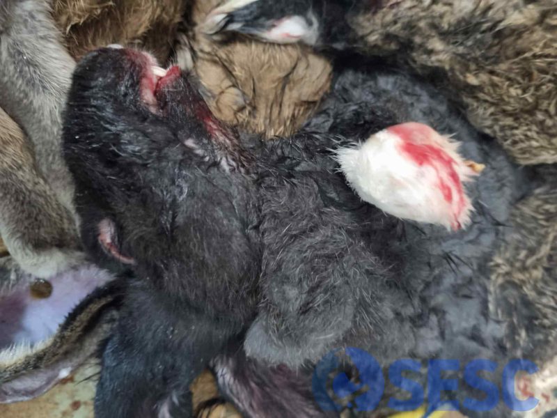
Animal found death in the antemortem examination, that presents blood in the nostrils, mouth and skin.
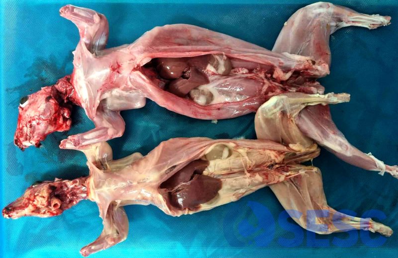
Pale and slightly icteric carcass (below).
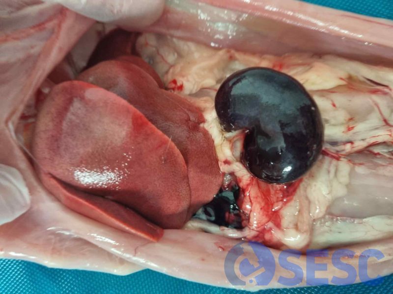
Rabbit carcass that presents an increase in size of the kidneys, with a dark coloration and a friable consistency (hemorrhagic appearance). Furthermore, the liver is pale and presents a lobular pattern.
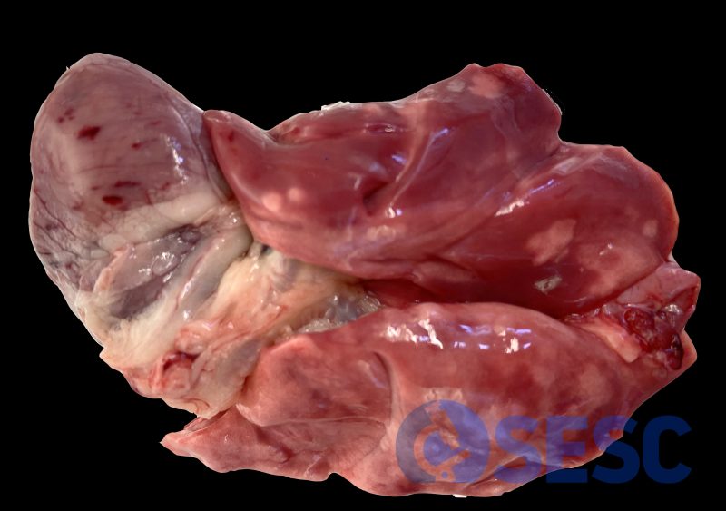
Diffusely reddish lungs and presence of petechiae in the epicardium.
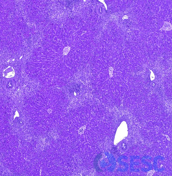
Histological section of the liver with necrotizing periportal to periacinar necrotizing hepatitis. Due to affecting periportal areas (in a bridging pattern) it is readily seen at low magnification as an intensification of the lobulillar pattern.
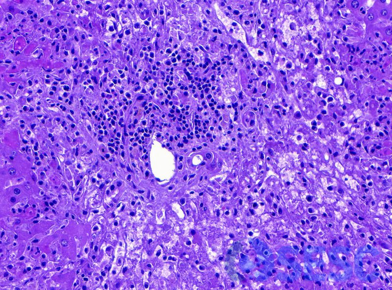
Hepatic lesion at higher magnification. The hepatocytes display tumefaction, cytoplasmic rarefaction (hydropic degeneration) and necrosis, as well as occasional presence of fibrin and mixt inflammation.
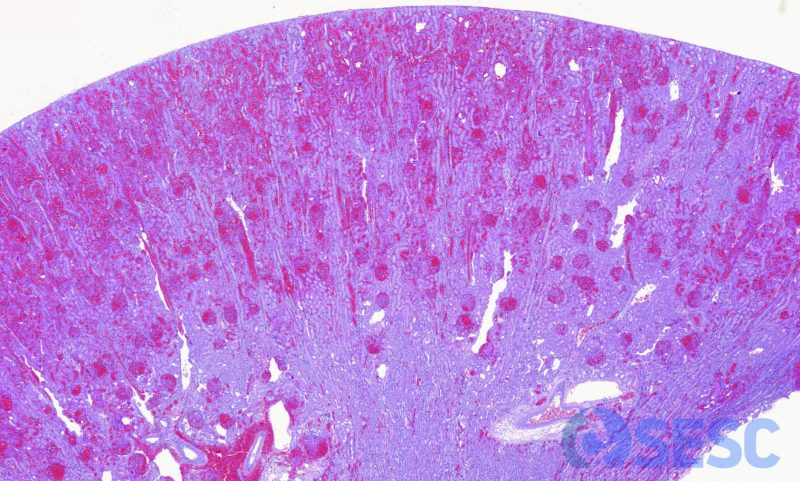
Histological section of the kidney at low magnification. Multifocal to coalescing haemorrhages are readily seen.
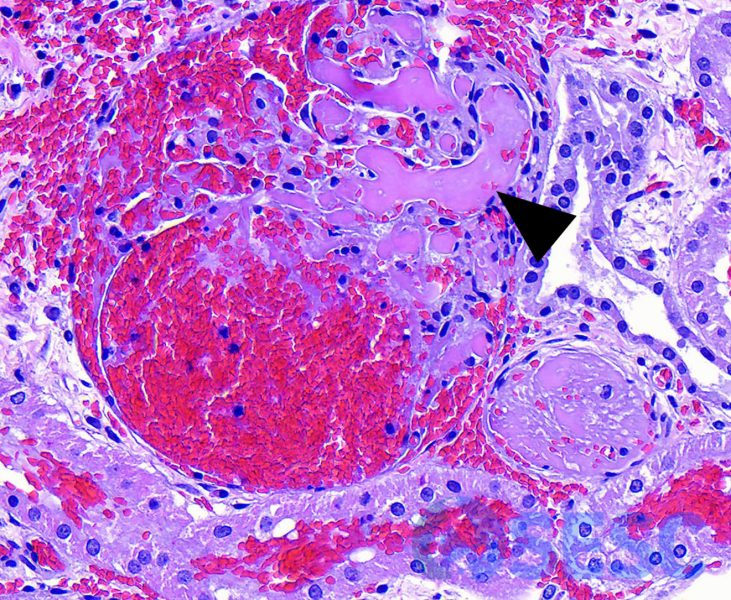
Kidney at higher magnification. Glomeruli shows diffuse thrombosis of its glomerular capillaries and haemorrhage into Bowman’s capsule.
