Any given day in a rabbit slaughterhouse (2023)
Last July we organized a practical workshop on identification and description of injuries for official slaughterhouse veterinarians. For this, we collected condemned viscera and carcasses from several rabbit slaughterhouses, which were the subject of discussion during the session.
In this entry, a couple of lesions are documented from which samples were also collected in formalin for histological study. (AC)
SESC pathologists and collaborators, Natàlia Majó and Carlos López-Figueroa, led the presentation of rabbit SESC cases.
CASE 1
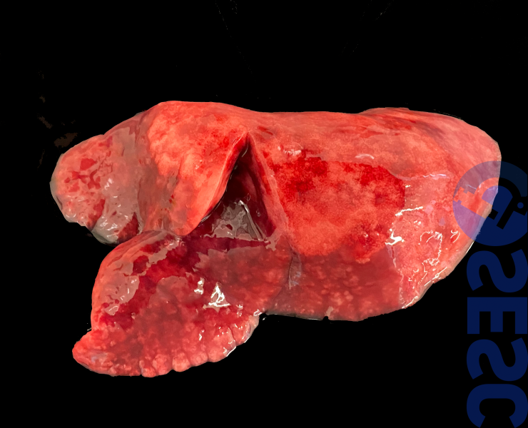
Rabbit right lung. A well-defined redness and increase in consistency is observed, affecting the cranioventral regions of the lung lobes. When cutting a portion of consolidated lung tissue, it sinks in water, while the rest of the tissue floats.
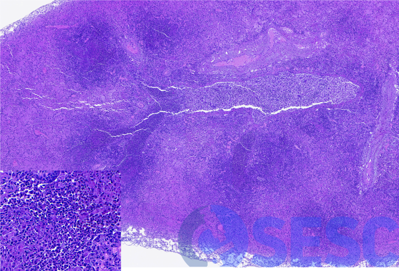
Histologically, pulmonary consolidation was confirmed, that is, the obliteration of the air space due to the presence of exudate inside the alveoli and bronchioles. In this case you can see how the airways had a denser exudate (that is, more basophilic, due to the presence of nuclei), while the alveoli had fewer inflammatory cells, and abundant edema. At higher magnifications (insert) it can be seen that the exudate is composed mainly of heterophils and - to a lesser extent - of mononuclear cells.
Catarrhal-suppurative bronchopneumonia is very similar between species, both for the lesions observed and for its pathogenesis. It is always the result of a noxa (almost always bacteria) that gains access to the lungs through the airways. Any agent that is able to interfere with the functioning of the mucociliary barrier will facilitate its development. The agents most commonly isolated in rabbits are P. multocida (isolated in this case) and B. bronchiseptica.
CASE 2
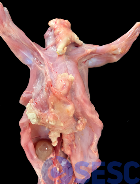
Rabbit carcass, in which accumulations of suppurative-caseous material are observed in the subcutaneous and muscular tissue, with multifocal and irregular distribution; as well as abscess formation.
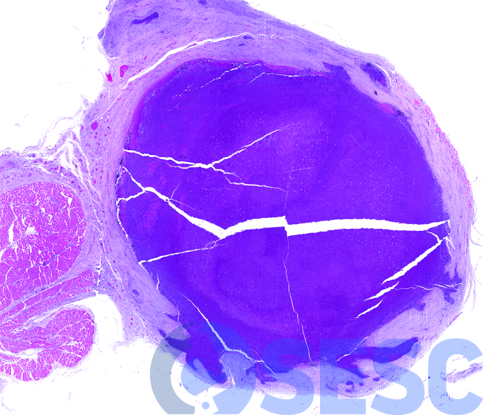
Histologically, the presence of multiple abscessing lesions (sometimes irregular), composed of very abundant viable and degenerated heterophils, delimited by a thick fibrous capsule, is confirmed.
These subcutaneous lesions (sometimes they can be found disseminated in the thoracic or abdominal cavities) are the product of bacterial infections, almost always Pasteurella multocida, which gain access to the subcutaneous tissue for example through small wounds or injections.
