

Infection with peste des petits ruminants virus (PPR virus infection)
Peste des petits ruminants is a disease affecting sheep and goats. It may also cause subclinical infection in cattle, camelids and pigs, but these species do not transmit the virus. Transmission occurs through direct contact with infected animals, fomites, inhalation or ingestion. PPR is listed by the World Organisation for Animal Health (WOAH, formerly OIE) as a notifiable disease and is classified as a Category A disease under Commission Delegated Regulation (EU) 2020/687.
The causative agent is an RNA virus of the family Paramyxoviridae, genus Morbillivirus, closely related to canine distemper virus, measles virus in humans, and rinderpest virus in cattle.
The virus typically enters via the respiratory (aerosols) or oral route. Initial replication occurs in the tonsils and mucosa-associated lymphoid tissue (MALT), followed by a primary viraemia. The virus infects monocytes, macrophages and T lymphocytes, leading to lymphoid depletion and severe immunosuppression in lymphoid tissues (necrosis in lymph nodes, spleen, bone marrow and gut-associated lymphoid tissue – GALT). A secondary viraemia then disseminates the virus to target organs: respiratory, digestive, urogenital tract and skin. Clinical signs include fever, erosive-ulcerative lesions in the oral cavity and pharynx, diarrhoea, pneumonia and death. The disease does not produce vesicles; lesions result from direct viral replication and necrosis of epithelial cells. Post-mortem findings often include tracheitis and fibrino-ulcerative enterocolitis.
PPR is endemic in the Middle East, South Asia and Africa. It was first reported in the European Union in 2018 in Bulgaria, and later in 2024 in Greece, Romania and again in Bulgaria, and in 2025 in Hungary.
Differential diagnoses include: bluetongue, foot and mouth disease, caprine infectious rhinotracheitis (caprine herpesvirus type 1), enterotoxaemia (Clostridium perfringens, particularly type D, due to diarrhoea), and border disease virus (BDV).
In cases with compatible lesions, diagnosis relies on histopathology (presence of syncytial cells with eosinophilic intracytoplasmic or intranuclear inclusions), immunohistochemistry, and RT-PCR for viral genome detection. Preferred samples are lung, spleen, mesenteric and mediastinal lymph nodes, ulcerated mucosa and intestine. The Spanish National Reference Laboratory for PPR is the Central Veterinary Laboratory (LCV) in Algete. (EV)
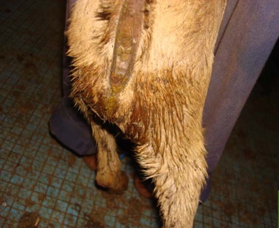
Goat, ante-mortem. Diarrhoea (soiled perianal region). Source: OMSA

Goat. Ante-mortem. Conjunctival congestion. Source: Balamurugan et al 2014
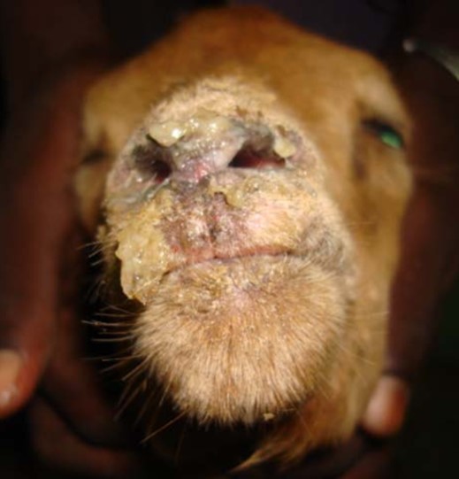
Goat. Ante-mortem. Mucopurulent nasal discharge. Source: OMSA
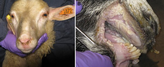
Sheep. Mucopurulent nasal discharge. Goat. Necrosis/erosions of the oral mucosa (no vesicles). Source: Truong et al 2014.
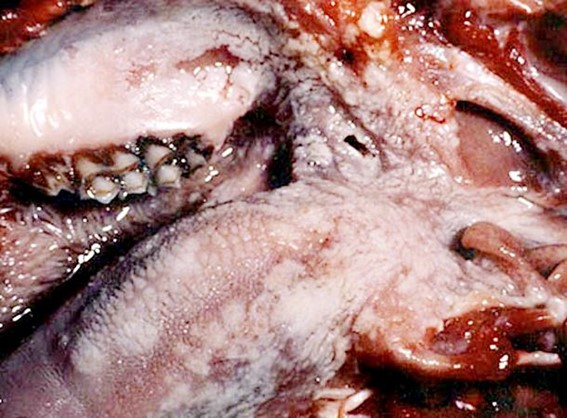
Goat. Epithelial necrosis (whitish areas) of the tongue and pharynx. Source: USDA-APHIS
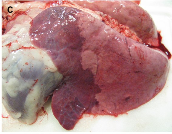
Goat. Secondary bronchopneumonia (due to immunosuppression). Source: Truong et al 2014

Goat. Zebra colon (longitudinal congestive stripes). Source: Balamurugan et al 2014 .

Goat. Colon with longitudinal congestive markings. Source: OMSA

Goat. Depression and reddening of Peyer’s patches. Source: Truong et al 2014.