

African swine fever (ASF) lesions in wild boar carcasses
At the end of 2025, an outbreak of African swine fever (ASF) was declared in wild boar in Catalonia and is currently still active. To date, all detected cases have been confined within a 6 km radius of the first carcasses found. Nevertheless, a high level of vigilance is required in case the outbreak spreads beyond this area, as the introduction of the virus into domestic pig populations would have very serious consequences.
This post presents some of the most characteristic lesions observed in wild boar necropsied in the context of this outbreak, with the aim of facilitating their recognition. If such lesions are detected in wild boar carcasses at game meat processing establishments, suspicion should be raised and the findings must be immediately notified to the competent authority (DARPA).
It should be recalled that ASF may cause acute death, sometimes in the absence of visible lesions. However, subacute forms of the disease may show haemorrhagic lesions in animals that are still alive and therefore potentially hunted. These lesions must be correctly identified and differentiated from haemorrhages caused by hunting projectiles, which are typically focal and associated with blood resorption in the regional lymph nodes. (EV)

Enlargement and reddening of the submandibular lymph nodes (sectioned in the image). The reddening is due to haemorrhagic lesions within the lymphoid tissue, showing an irregular appearance. This finding must be differentiated from blood resorption, for example when the wild boar has been shot close to the head. In such cases, an accumulation pattern in subcapsular and peritrabecular sinuses is observed (see examples here and here).
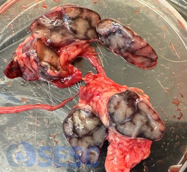
Example of a cut lymph node from a healthy wild boar, without ASF, hunted by a hunter, in which reabsorption of blood can be observed in the subcapsular and peritrabecular sinuses, probably due to reabsorption of blood from hemorrhages caused by the shot. This should not be confused with hemorrhages of the lymphoid tissue itself. Over time, due to autolysis and also in frozen tissues, hemoglobin may diffuse and the tissue may show a more reddish coloration.
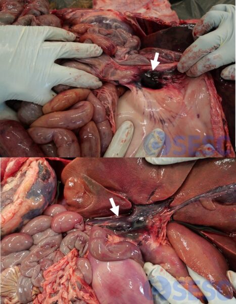
Enlargement and haemorrhagic appearance of the gastrohepatic lymph node (arrow). This finding is highly characteristic of African swine fever in both wild boar and domestic pigs. Images from two different animals are shown.
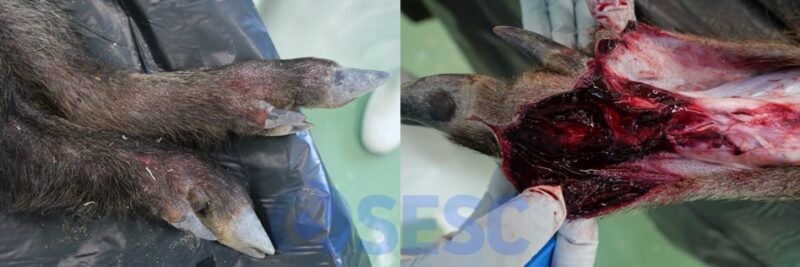
In some cases, erythematous skin lesions are observed. After skin removal, subcutaneous haemorrhages can be identified.
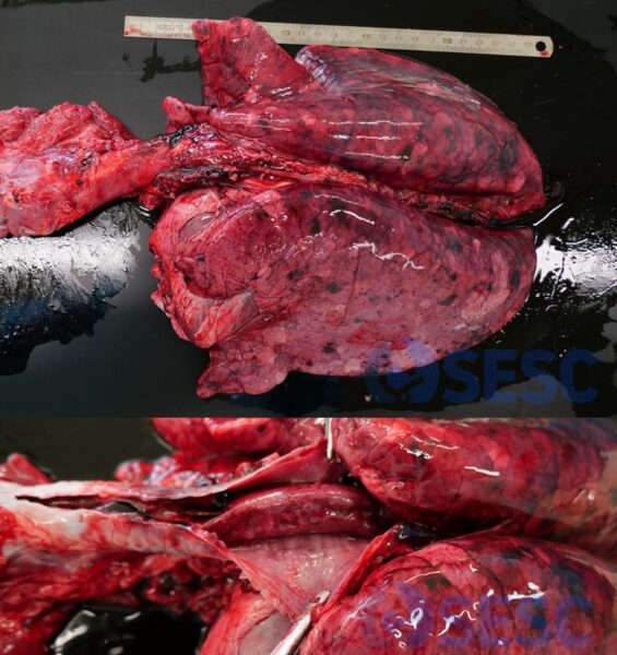
After removal of the lungs from the thoracic cavity, lack of pulmonary collapse is observed due to pulmonary oedema. In addition, the lungs are congested (reddened) and show multifocal haemorrhages (petechiae and ecchymoses). In the lower part of the image, opening of the trachea reveals the presence of a blood clot within the airways.
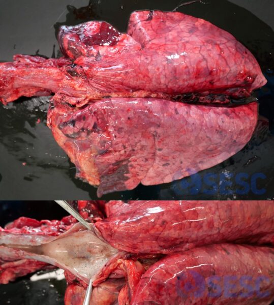
Another image of lungs failing to collapse after removal from the thoracic cavity, with haemorrhages and distension of the interlobular septa (interstitial oedema). Upon opening the trachea (lower part of the image), the presence of foam within the airways is observed (alveolar oedema).
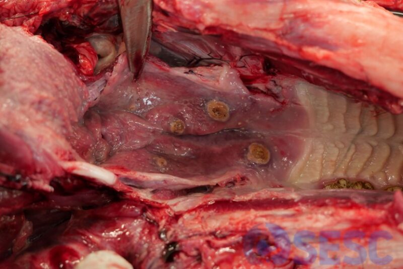
Foci of necrosis in the palatine tonsils. This lesion is not described as a typical lesion in ASF (whereas it is in CSF); however, we have observed these lesions in a few wild boar affected by the current outbreak.
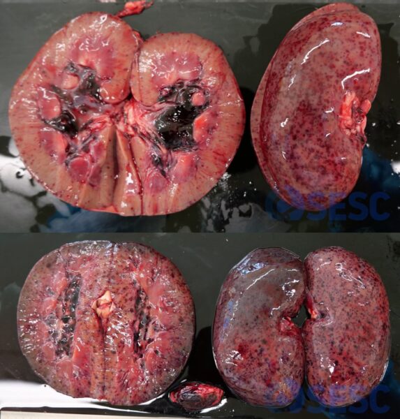
Multifocal renal haemorrhages (petechiae), mainly distributed in the renal cortex and renal pelvis. Images from kidneys of two different animals are shown. This is a fairly characteristic lesion and is unlikely to be observed as a consequence of gunshot wounds. The renal lymph nodes are also reddened (haemorrhagic).
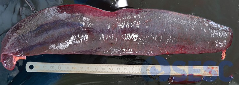
Splenomegaly, or enlargement of the spleen, is also a characteristic finding in African swine fever, although it is not specific to this disease.