African Swine Fever lesions
The images were obtained from experimental infections carried out at the level 3 biocontainment unit in CReSA, within the framework of research projects on this disease and with the approval of the animal experimetnation ethics committee at the UAB. It's an infection with ASF virulent strains, similar to those circulating nowadays in Eastern Europe and causing acute lesions, different than the attenuated strains, that cause chronic lesions, and which will likely appear later in time.
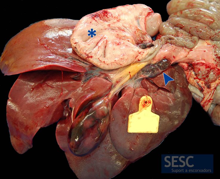
Hemorrhage of the gastrohepatic lymph node (arrow), this one is a very ASF characteristic lesion. The gallbladder shows areas of hemorrhage in the wall. In the gastrointestinal tract serosa petechiae can be obserevd, such as the stomach of the image (asterisk).
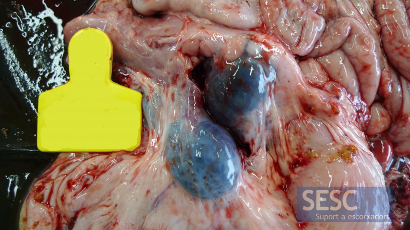
Another image of the gastrohepatic hemorrhagic lymph node. This is one of the lesions observed more consistently in animals inoculated with ASF.
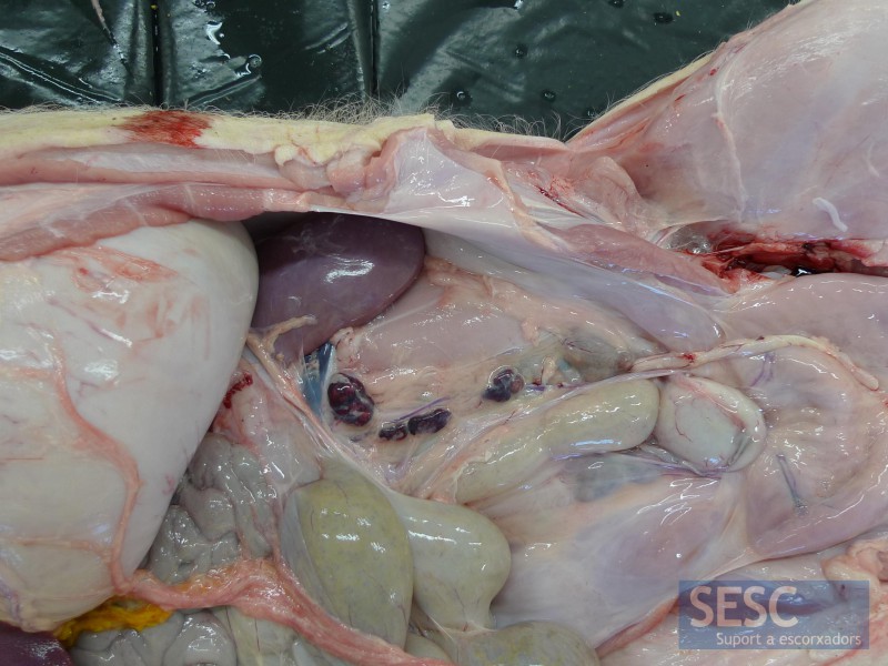
Renal lymph nodes can also often be hemorrhagic.
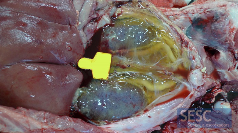
In more severe cases perirenal edema is observed, in the image we had removed the peritoneum. The kidneys have multiple petechial hemorrhages.
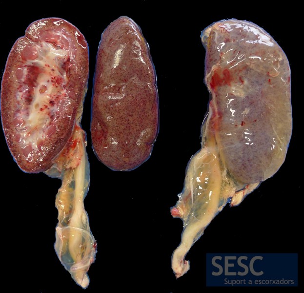
Petechial hemorrhages (<3 mm in diameter) of miliary distribution in the cortex and renal pelvis.
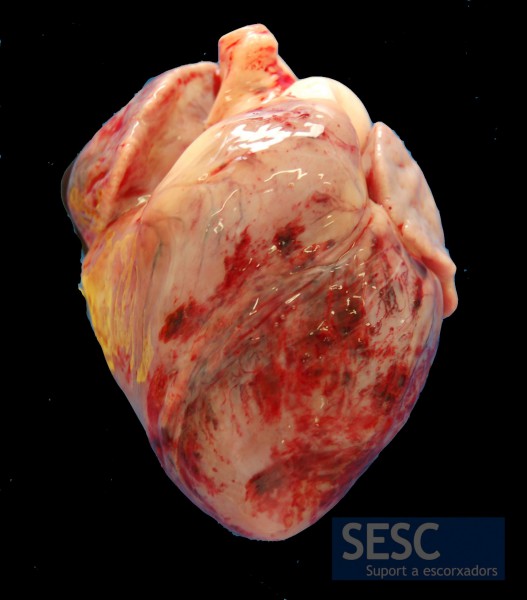
Epicardial hemorrhages.
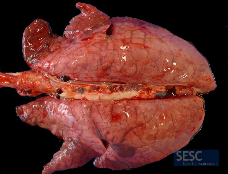
Intense pulmonary edema, evidenced by the lack of collapse of the lungs with evident interstitial edema between lung lobules. This lesion is not specific to ASF.
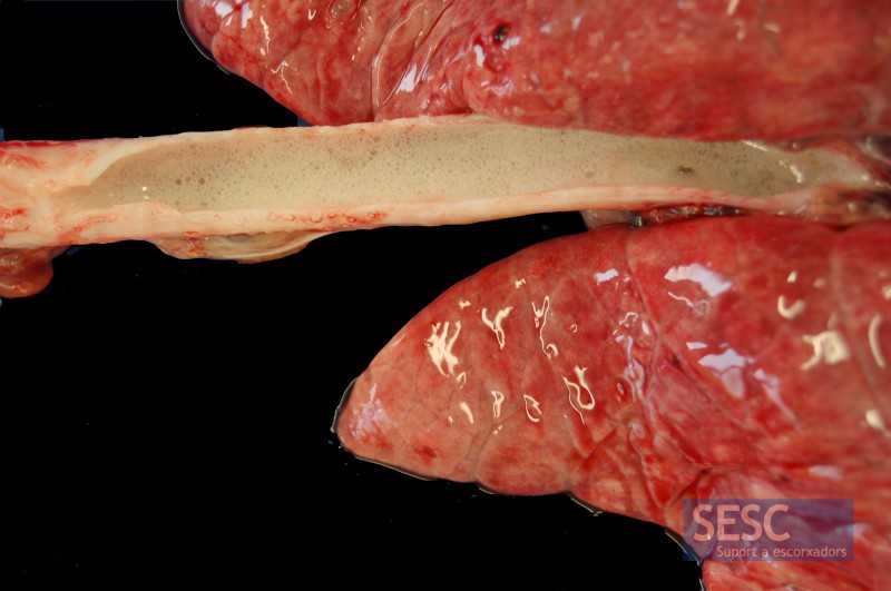
Marked pulmonary edema, evidenced by the presence of foam inside the trachea (open longitudinally). This lesion is not specific to ASF.
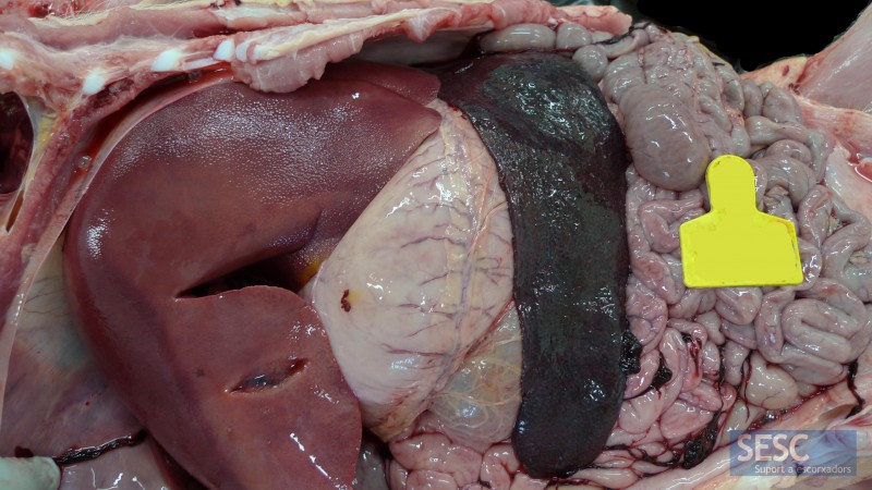
Marked increase in spleen size (splenomegaly). This lesion is not specific for ASF.
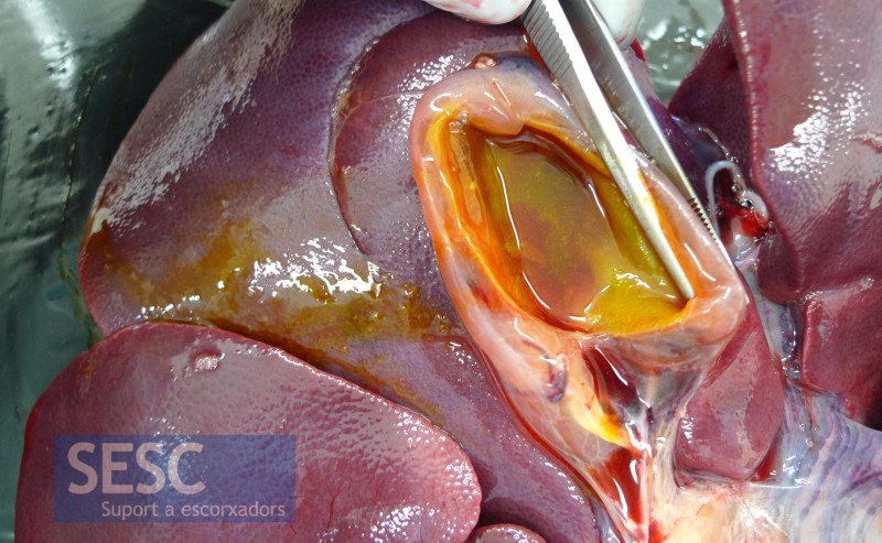
Another common lesion is edema of the gall bladder wall.
