Ammonium urate concretions in the splenic capsule of a calf
In a 12-month-old, male, Montbeliard breed, calf carcass multiple sand-like, roughly granular, whitish, spherical concretions of crystalline appearance were observed attached to the spleen capsule.
When processing the tissue for histopathology the mentioned structures disappeared (probably dissolved in the fixative). Histologically, there was no inflammatory reaction but the presence of multiple, mostly gram-negative, bacteria was observed.
The study by infrared spectroscopy of the structures reveals that these are ammonium urate calculi.
The findings described in this case have an uncertain origin:
- On the one hand the absence of inflammation would suggest that the presence of bacteria could be a postmortem contamination of the tissue (the microbiological culture allows to identify: Klebsiella, Proteus, Staphylococcus and Brevibacterium).
- On the other hand there are no descriptions in calves of the formation of urate crystals or calculi. One hypothesis would be that it was a visceral gout, secondary to hyperuricemia, but this pathology is not described in ruminants, nor would it explain why only the spleen is affected.
If you have found similar lesions, we invite you to comment in the comment section at the end of the post.
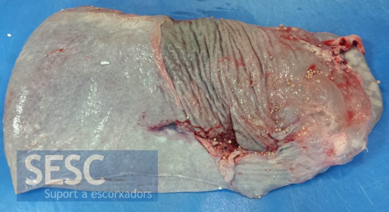
Multiple spherical, solid, whitish concretions adhered to the splenic capsule.
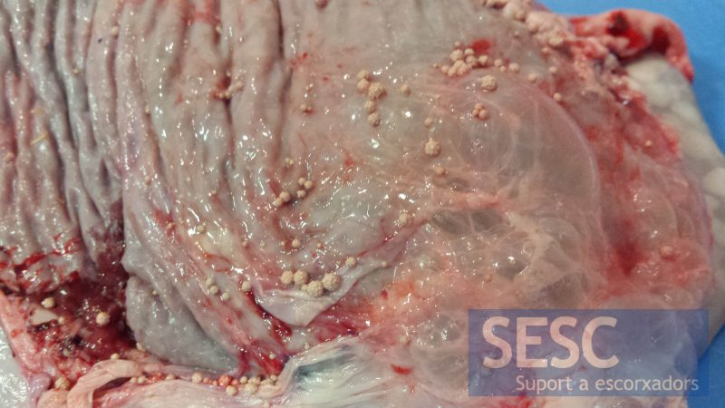
Detail of the concretions.

2 comment(s)
Comment by Scott Reed in LinkedIn’s Veterinary Pathology group:
I have not seen these in cattle, but I have seen fecoliths in megacolon cats that were presumably associated with previous colon rupture and healing as well as struvite uroliths attached to the mesentery of a dog presumably associated with previous bladder rupture – Osborne’s group at U Minn have described spontaneous bladder healing after rupture in dogs and cats and even suggest it as a possible treatment modality. Any scars evident on the urinary bladder, renal pelvis, or ureters?
Hello Scott, thanks for your comment! We considerd that option (bladder or urinary tract rupture) still we haven’t found descriptions of such urinary calculi in cattle.
Only the spleen was submitted and no other macroscopic changes were reported.