Mystery Dermatitis. Volume 2.
In a male, cross breed, 6 months old, pig carcass multiple, multifocal, erythematous lesions were observed some of which with concentric circles morphology. Generalized lymphadenopathy was also observed with increased size and redness of the lymph nodes.
Samples submitted for histopathology were taken after scalding, precluding an appropriate assessment of the histological lesions in the epidermis and the superficial dermis due to the artifacts caused by this process. In the deeper dermis, perivascular and periadnexal dermatitis was observed. The lymph nodes were hyperplastic and with abundant presence of eosinophilic polymorphonuclear leukocytes. The kidneys showed no obvious lesions.
Despite having a very characteristic lesional pattern the aetiology could not be determined. A while ago another case of Mystery dermatitis in a pig carcass was submitted, of which the ethiology could not be determined, in that case the lesions were radial.
The differential diagnosis could include:
- Porcine Dermatitis and nephropathy syndrome (PDNS): histopathological study of the kidneys ruled this one out, which was the main suspicion of the inspectors who submitted the case.
- Pityriasis rosea: swine juvenile psoriasiform pustular dermatitis. It seems one of the most probable differentials.
- Dermatophytosis (ringworm).
- Erysipelas: despite the characteristic rhomboid morphology of lesions was not observed.
In human medicine figured erythemas are described, some of which (eg erythema annulare centrifugum) are reminiscent of images we see in this pig carcass. The causes can be varied: from primary disease of the skin (tinea, urticaria ...) to systemic diseases (bacterial infections, neoplasms ...) presenting this lesion.
Again, if any of you have seen such a lesion, we encourage you to discuss this in the comments section at the end of this post.
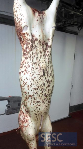
Lesions on the ventral aspect of the carcass.
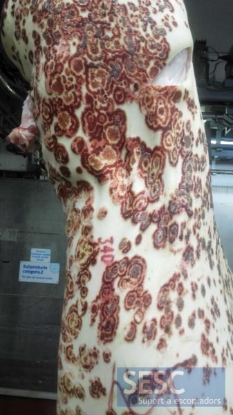
Detail of the lesions on the dorsal aspect, showing a particular concentric pattern.
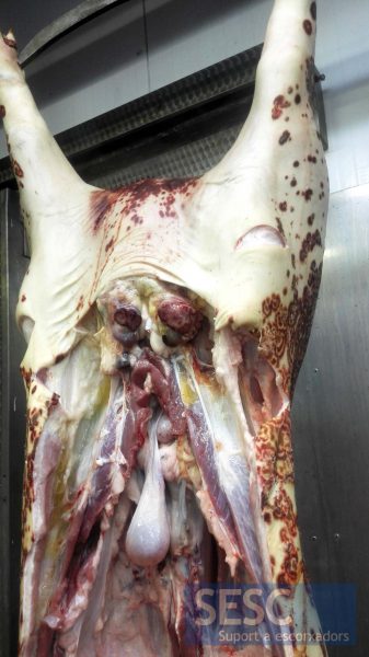
Increased size and redness of the superficial inguinal lymph nodes.

4 comment(s)
A mí ese halo hiperémico en forma de onda de agua me sugiere una infección fúngica.
Perhaps autoimmune such as erythema multiforme (Stevens Johnson Syndrome), granuloma annulare, etc. One would require a good histologic picture obviously to be sure of the target of the reaction. Vasculitis versus perivascular infiltrate is an important differentiation obviously.
Comment from Elena Riccardi in LInkedIN Veterinary pathology group:
I was thinking about an infectious cause of the lesion (viral disease, PDNS) but I guess the animal was not pyrexic. Vasculitis?
Comment from David K. Meyerholz in LInkedIN Veterinary pathology group:
The macroscopic and microscopic lesion pattern, distribution on the carcass, and age of the animal would suggest dermatophytosis (e.g. Microsporum and Trichopyton spp.) as a leading differential diagnosis. Gross lesions identical to these have been reported for dermatophytosis infection in pigs. Other reasonable considerations could include PDNS and Pityriasis rosea, but the age, pattern and/or distribution of lesions in this case would be an atypical presentation.